|
Olympus BioScapes Digital Imaging Competition 2012
|
||
|
1st Place
Ralph Grimm
Jimboomba, Queensland, Australia
Specimen: Colonial rotifers showing eyespots and corona, magnification 200x - 500x.
Technique: Differential interference contrast
Microscope images forge an extraordinary bond between science and art, said Hidenao Tsuchiya, Olympus America's Vice President and General Manager for the Scientific Equipment Group. We founded this competition to focus on the fascinating stories coming out of today's life science research laboratories. The thousands of images that people have shared with the competition over the years reflect some of the most exciting work going on in research today – work that can help shed light on the living universe and ultimately save lives. We look at BioScapes and these beautiful images as sources of education and inspiration to us and the world
2nd Place
Dr. Arlene Wechezak
Anacortes, Washington, USA
Specimen: Red algae Scagelia, showing reproductive tetraspores and golden diatoms.
Technique: Darkfield
3rd Place
Dr. Igor Siwanowicz
HHMI Janelia Farm Research Campus
Ashburn, Virginia, USA
Specimen: East-coast US fern, Polypodium virginianum, showing a cluster of spore-filled sporangia and specialized protective hairs called paraphyses.
Technique: Confocal microscopy
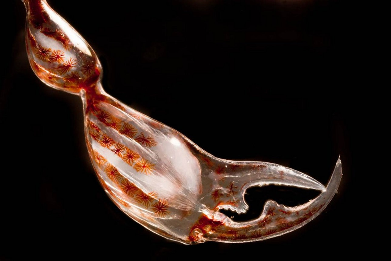
4th Place
Dr. Christian Sardet and Mr. Sharif Mirshak
The Plankton Chronicles Project
Villefranche-sur-Mer, France, and Montreal, Quebec, Canada
Specimen: Claw of crustacean amphipode Phronima sp. Muscles and rows of pigment cells (melanocytes) are visible.
Technique: Darkfield
5th Place
Mr. Rogelio Moreno Gill
Biomarcell Station Zoologique
Panama City, Panama
Specimen: Unicellular green alga Micrasterias from lake sample. 22 stacked images.
Technique: Differential interference contrast
6th Place
Mr. James Nicholson
NOAA/NOS/NCCOS Center for Coastal Environmental Health & Biomolecular Research
Fort Johnson Marine Lab, Charleston, SC, USA
Specimen: Live mushroom coral Fungia sp. Close-up of mouth during expansion.
Technique: Autofluorescence
7th Place
Dr. Christian Klämbt and Dr. Imke Schmidt
University of Münster, Münster, Germany
Specimen: Beta-tubulin expression of a Drosophila third instar larval brain, with attached eye imaginal discs.
Technique: Confocal microscopy
8th Place
Mr. Edwin Lee
Carrollton, Texas, USA
Specimen: Henbit (Lamium amplexicaule) stamens anthers and filaments.
Technique: Phase contrast
9th Place
Mrs. Sahar Khodaverdi
University of Tabriz
Tabriz, East Azerbaijan, Iran
Specimen: Delphinium seed. The image was acquired from multiple Z-stacked images.
Technique: Epi-fluorescence
10th Place
Mr. Charles Krebs
Issaquah, Washington, USA
Specimen: Butterfly "Prola Beauty" (Panacea prola) wing scales, 200X.
Technique: Diffused reflected illumination
Olympusbioscapes |
||
|
|
|
|
| ← Previous picture Next picture → | ||
|
Military Woman Gallery
Must See Places |
||
|
|
||