|
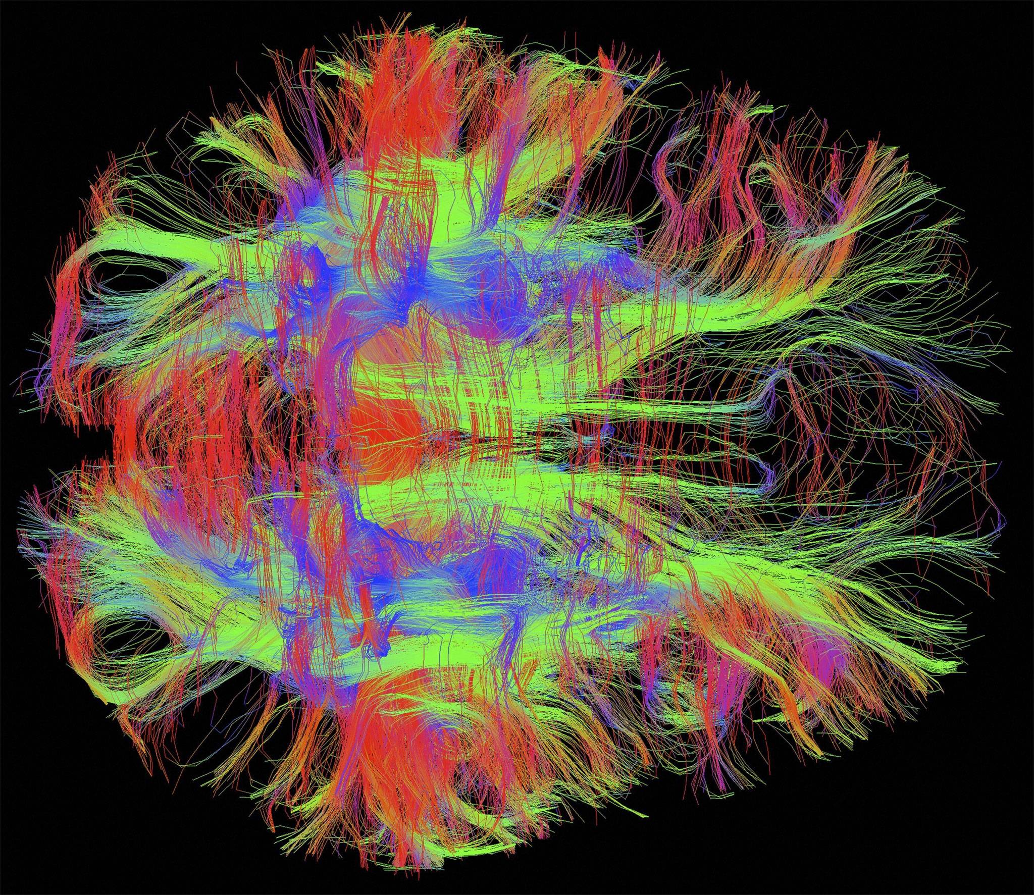
Bird's eye (axial) view of nerve fibres in a normal, healthy adult human brain. Brain cells communicate with each other through these nerve fibres, which have been visualised using diffusion weighted magnetic resonance imaging (DWI MRI). Diffusion weighted imaging is a specialised type of MRI scan which measures water diffusion in many directions in order to reconstruct the orientation of the nerve fibres. Since these images are in 3D, colours have been used to represent the direction of the fibres: blue is for fibres travelling up/down, green for front/back, and red for left/right. These patterns of connectivity in the brain are being used to study brain development and developmental disorders such as dyslexia. (Photo by Zeynep M. Saygin/McGovern Institute/MIT/Wellcome Images)
|