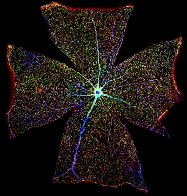
Surface of a mouse retina. It looks like a delicate floral structure. In fact it is the surface of a mouse retina. Produced by combining more than 400 images captured through a technique known as confocal microscopy, this digital reconstruction shows the retina’s blood vessels in blue while the red and green highlight cells known as astrocytes. These star-shaped cells are involved in myriad crucial processes that support and maintain neurons. Scientists hope that by understanding what astrocytes are up to as the retina deteriorates, they might find new ways to treat vision loss. (Photo by Gabriel Luna/Neuroscience Research Institute/University of California/Wellcome Images)
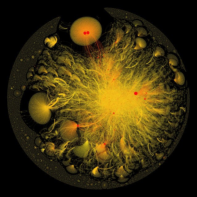
#breastcancer Twitter connections. A stunning, vibrant infographic, this image was created by collecting and analysing tweets which used the hashtag #breastcancer. The connections between Twitter users were then explored, giving rise to this intricate and elaborate graphic in which the dots represent users and the lines reflect their interactions. (Photo by Eric Clarke, Richard Arnett and Jane Burns/Royal College of Surgeons in Ireland/Wellcome Images)
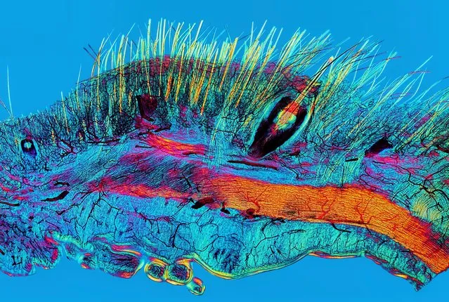
Cat skin and blood supply. This image, captured from a Victorian microscope slide using polarised light microscopy shows, as the title reveals, cat skin and its blood supply. Fine hairs and thicker whiskers are shown in yellow while blood vessels are shown in black. More than 40 images were combined to produce the final colourful image. (Photo by David Linstead/Wellcome Images)
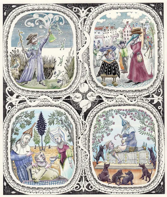
Caricatural medieval medical practitioners. This digital illustration is darkly humorous, depicting a range of figures from an alchemist shown triumphantly clutching a flask to a surgeon pulling what looks like a string of sausages from the stomach of an alarmed-looking patient. (Photo by Madeleine Kuijper Illustraties/Wellcome Images)
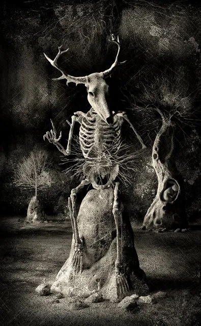
Stickman – The Vicissitudes of Crohn’s. The overall winner of this year’s Wellcome Image Awards is this eerie depiction of Crohn’s disease. The scene is a digital image which captures the artist’s own experience of the condition. Thought to be caused by inflammation of the digestive system, Crohn’s disease often involves abdominal pain and weight loss. With the character Stickman the artist reflects such symptoms, but captures signs of hope, too, not least in the hare nestled snugly in the trunk of the tree. (Photo by Spooky Pooka/Wellcome Images)
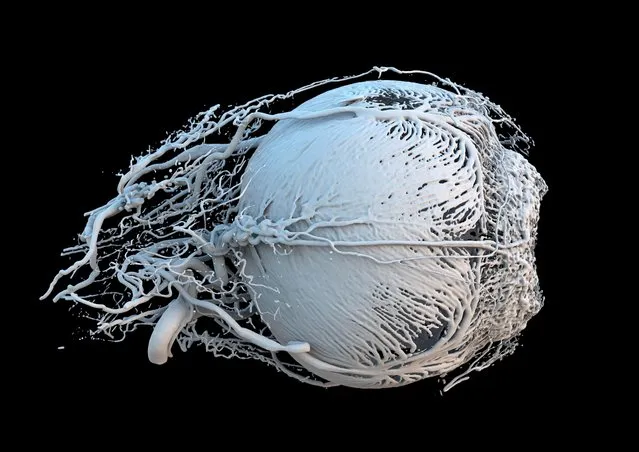
Vessels of a healthy mini-pig eye. Produced from CT scans and 3D printing, this is a model of the blood vessels of the eye of a miniature pig. A small as 0.02 mm in diameter, the vessels are vital in the delivery of nutrients to muscles which change the size of the pupil, constricting or dilating its size to regulate how much light enters the eye. (Photo by Dr Peter M Maloca/OCTlab/University of Basel and London Moorfields Eye Hospital; Christian Schwaller, Ruslan Hlushchuk/University of Bern and Sébastien Barré/Wellcome Images)
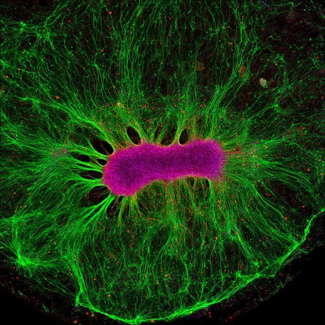
Brain-on-a-chip. Dazzling in green and magenta this image shows the nerve fibres (in green) produced by neural stem cells (in magenta) as they grow on a synthetic gel. Captured by a technique known as confocal microscopy, the image is part of research shedding light on how tinkering with the environment can affect the way in which nerve fibres grow. (Photo by Collin Edington and Iris Lee/Massachusetts Institute of Technology/Wellcome Images)

Two young boys in rural Nicaragua. This photograph highlights the devastating impact of chronic kidney disease in the Nicaraguan town of Chichigalpa. These brothers have lost cousins and uncles to a form of the disease which is thought to be linked to heavy labour in hot temperatures. (Photo by Joshua Mcdonald/Wellcome Images)
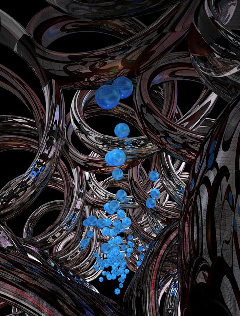
Synthetic DNA channel transporting cargo across membranes. This digital illustration shows the artist’s impression of tube-like channels in a cell membrane that have been constructed by researchers from DNA. The title of the piece references two components of the structure – the channel itself, shown in silver, and substances travelling through the channel, shown in blue. (Photo by Michael Northrop/Wellcome Images)
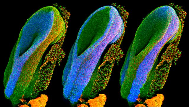
Developing spinal cord. This image shows a mouse’s neural tube – the structure from which the spinal cord forms during pregnancy. The different images highlight the three main tissue types in the embryo. (Photo by Gabriel Galea/University College London/Wellcome Images)
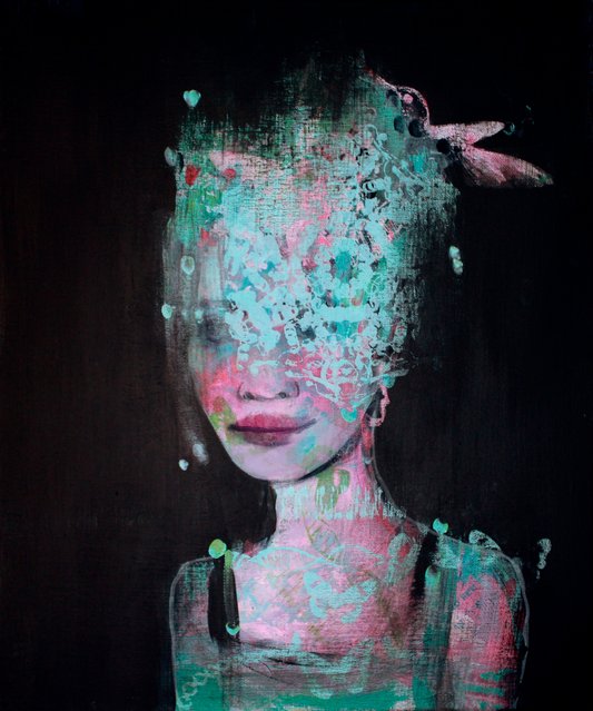
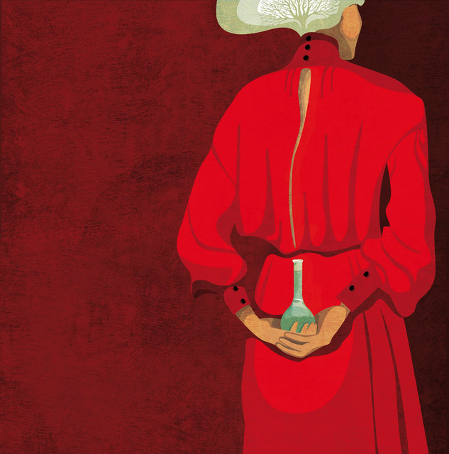
Hidden Learning. This painting draws on the experiences of women in science, in particular reflecting the myriad aspects of life that women keep to themselves while at work. (Photo by Chrysalis project/University of St Andrews/Wellcome Images)
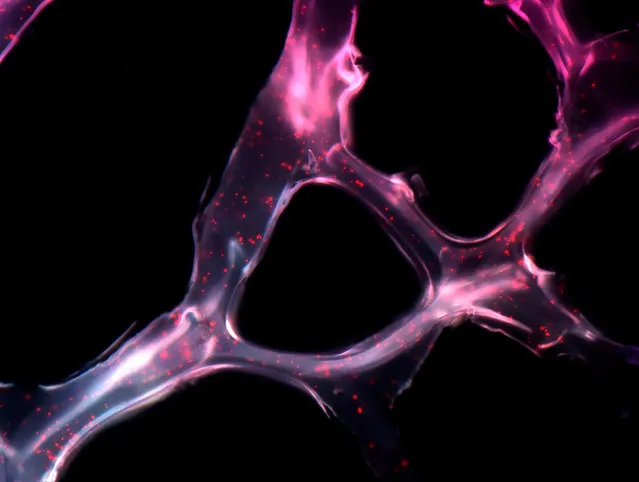
MicroRNA scaffold cancer therapy. This image shows a net-like structure, made from a synthetic polymer, that can be used to deliver two short, single-stranded genetic sequences known as microRNAs to cancer cells where they act to shrink tumours. The image was captured using scanning tunnelling microscopy – a technique that uses a beam of electrons to reveal the structure of objects. (Photo by João Conde. Nuria Oliva and Natalie Artzi/ Massachusetts Institute of Technology/Wellcome Images)
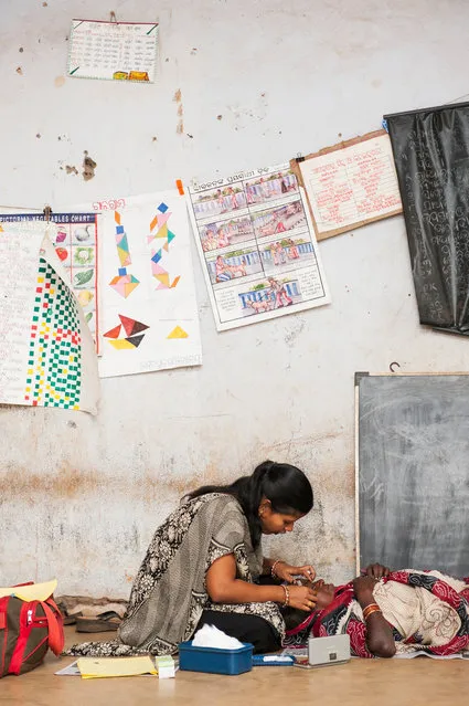
Patient receiving treatment during outreach eye screening in India. This photograph shows an eye doctor at work. At the time it was taken, the photographer was volunteering with a charity called Unite for Sight, which aims to improve eye health globally. (Photo by Susan Smart/Wellcome Images)
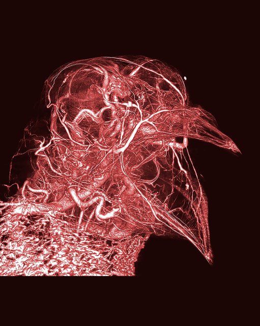
Pigeon thermoregulation. Bright, dramatic and intricate, this image reveals the network of blood vessels in a humble pigeon. Produced from CT scans, the image was made possible by the creator’s development of a novel contrast agent – a type of substance that helps to improve the visibility of structures within the body in medical imaging. (Photo by Scott Echols/Scarlet Imaging and the Grey Parrot Anatomy Project/Wellcome Images)
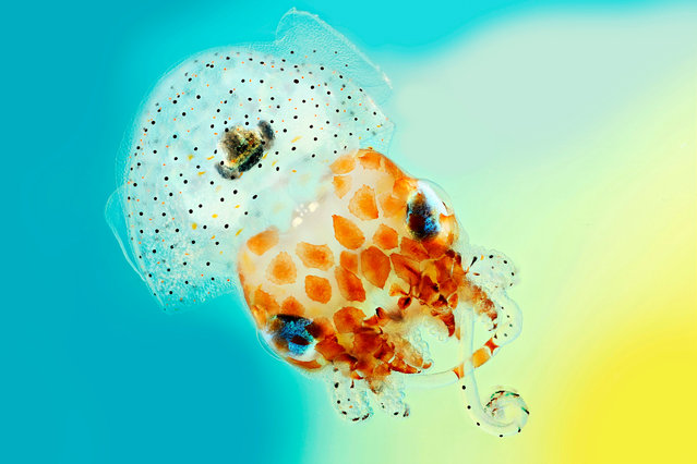
Hawaiian bobtail squid. This jaunty little Hawaiian bobtail squid was captured with close-up photography which produced a series of images that were then stitched together. The diminutive creature is a classic example of symbiosis: the squid offers a home and nutrients to light-producing bacteria, while the bacteria allow the squid to flit through the water without creating a telltale silhouette. (Photo by Mark R. Smith/Macroscopic Solutions/Wellcome Images)
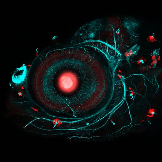
Zebrafish eye and neuromasts. This image was captured using confocal microscopy. The lens of the zebrafish is shown in red in the centre of the image, its nervous system is shown in blue, while the surrounding red structures are the neuromasts – cells which underpin the ability of fish to detect changes in the movement of the water. This in turn plays a role in all manner of behaviours including helping fish pick up the movements of prey. (Photo by Ingrid Lekk and Steve Wilson/University College London/Wellcome Images)
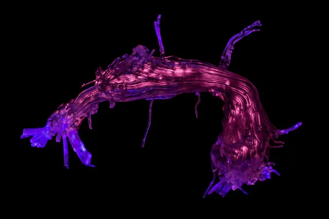
Language pathways of the brain. This colourful image shows a 3D model of an arched band of white matter in the brain, known as the arcuate fasciculus, which connects two regions of grey matter that are involved in speech and language. The model was created using 3D printing, with the colours produced by illumination, and was based on data collected through a type of magnetic resonance imaging. (Photo by Stephanie J. Forkel and Ahmad Beyh/Natbrainlab/King’s College London/Wellcome Images)
Rita Levi-Montalcini. A striking portrait, this digital image depicts the late neurobiologist Rita Levi-Montalcini. A medical graduate in Italy in the late 1930s, she was forced to work in secret on her early research as a result of Mussolini’s antisemitic laws. She later moved to the US and went on to discover nerve growth factor, a breakthrough that scooped her half of the Nobel Prize for Physiology or Medicine in 1986. (Photo by Daria Kirpach/Salzman International/Wellcome Images)
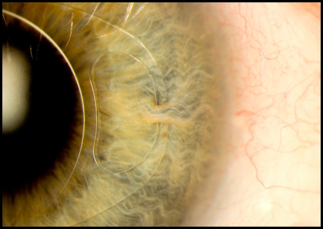
Intraocular lens “iris clip”. This photograph of the human eye shows a surgically implanted structure that can correct or restore vision. Different types of intraocular lenses can be used to tackle different conditions such as short-sightedness and even cataracts. This image was the winner of the Julie Dorrington Award for outstanding photography in a clinical environment. (Photo by Mark Bartley/Cambridge University Hospitals/NHS Foundation Trust/Wellcome Images)
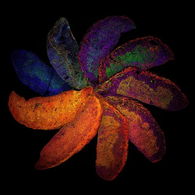
The Placenta Rainbow. Rich in oranges, blues and greens this is a vivid arrangement of mouse placentas. Captured through confocal microscopy, the image shows placentas taken from mice who have genetically different immune systems, highlighting how such differences can affect the structure of the placenta for example the development of blood vessels (shown in red) – an important consideration for conditions such as pre-eclampsia. (Photo by Suchita Nadkarni/William Harvey Research Institute/Queen Mary University of London/Wellcome Images)
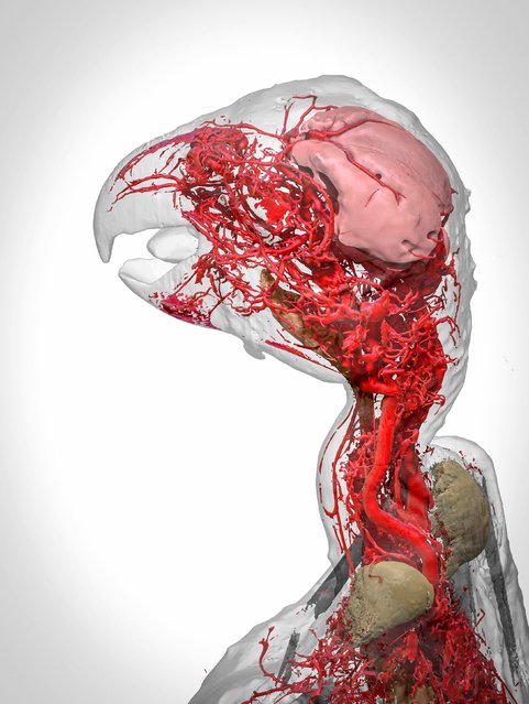
Blood vessels of the African grey parrot. A stunning view of the blood vessels of the African grey parrot, this 3D model was produced through computer modelling of 2D CT scans. The scans were captured by means of the same novel contrast agent developed by Scott Echols – creator of the earlier pigeon image. (Photo by Scott Birch/Scott Echols/Wellcome Images)
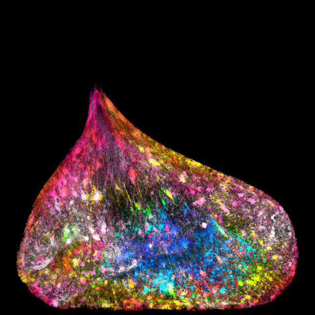
Unravelled DNA in a human lung cell. Multi-coloured and teardrop in shape, this was captured using super-resolution microscopy. It shows the nucleus of a lung cell which has been deformed due to a hitch during cell division. (Photo by Ezequiel Miron/University of Oxford/Wellcome Images)
17 Mar 2017 00:01:00,
post received
0 comments