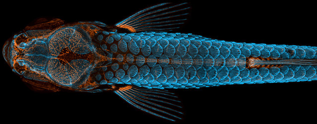
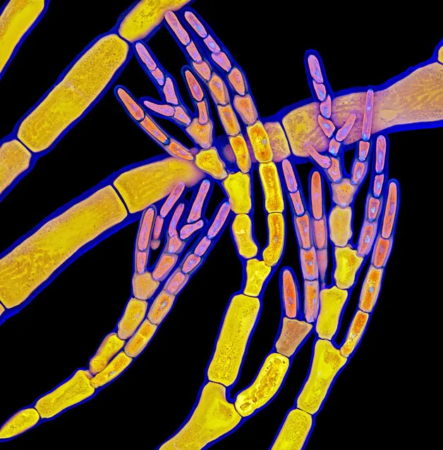
1st Place. Daniel Castranova, assisted by Bakary Samasa while working in the lab of Dr Brant Weinstein at the National Institutes of Health, took the top prize for his artfully rendered and technically immaculate photo of a juvenile zebrafish. The image is a dorsal view of the head of a fish with fluorescently “tagged” skeleton, scales (blue) and lymphatic system (orange), taken using confocal microscopy and image-stacking. 4X (objective lens magnification). (Photo by Daniel Castranova, Dr Brant Weinstein & Bakary Samasa/Nikon Small World Photomicrography 2020)
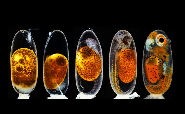
2nd Place. Embryonic development of a clownfish (Amphiprion percula) on days 1, 3 (morning and evening), 5, and 9, created using image-stacking. It shows the development, from hours after fertilisation (even with a pack of sperm cells being visible on top of the egg), until hours before hatching. The primary challenge was to create sharp focus stacking pictures while the embryo was alive and moving. (Photo by Daniel Knop/Nikon Small World Photomicrography 2020)
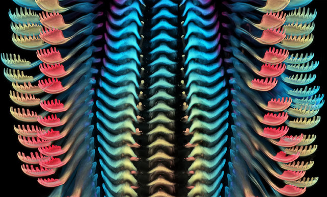
3rd Place. Small World veteran Dr Igor Siwanowicz captured this image of the tongue (radula) of a freshwater snail, using confocal microscopy. Confocal 40X (objective lens magnification). (Photo by Dr Igor Siwanowicz/Nikon Small World Photomicrography 2020)
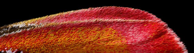
18th Place. Atlas moth wing. Image Stacking 10x (objective lens magnification). (Photo by Chris Perani/Nikon Small World Photomicrography 2020)
13th Place. Crystals formed after heating an ethanol and water solution containing L-glutamine and beta-alanine Polarized Light 4X (objective lens magnification). (Photo by Justin Zoll/Nikon Small World Photomicrography 2020)
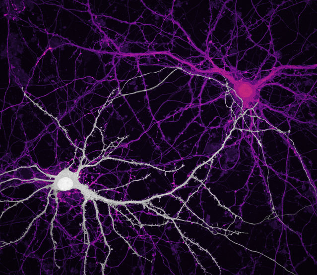
9th Place. Houston, Texas, US. Connections between hippocampal neurons (brain cells) Confocal 63X (objective lens magnification). (Photo by Jason Kirk & Quynh Nguyen/Nikon Small World Photomicrography 2020)
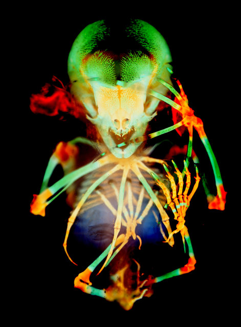
20th Place. Skeleton preparation of a short-tailed fruit bat embryo (Carollia perspicillata). Brightfield 1X (objective lens magnification). (Photo by Dr Dorit Hockman & Dr Vanessa Chong-Morrison/Nikon Small World Photomicrography 2020)
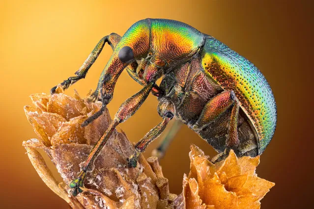
14th Place. Leaf roller weevil (Byctiscus betulae) lateral view. Image Stacking, Reflected Light 3.7X (objective lens magnification). (Photo by Özgür Kerem Bulur/Nikon Small World Photomicrography 2020)
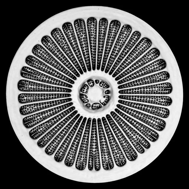
19th Place. Silica cell wall of the marine diatom Arachnoidiscus sp. Confocal 50x (objective lens magnification). (Photo by Dr Jan Michels/Nikon Small World Photomicrography 2020)
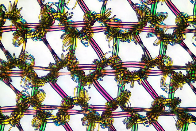
16th Place. Nylon stockings. Polarized Light 9X (objective lens magnification). (Photo by Alexander Klepnev/Nikon Small World Photomicrography 2020)
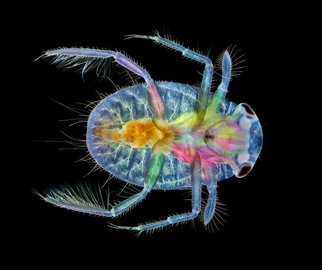
17th Place. Ventral view of an immature water boatman. Darkfield, Image Stacking, Polarised Light 4X (objective lens magnification). (Photo by Anne Algar/Nikon Small World Photomicrography 2020)
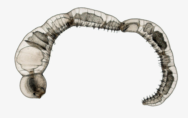
15th Place. Chain of daughter individuals from the asexually reproducing annelid species Chaetogaster diaphanus. Brightfield 5X (objective lens magnification). (Photo by Dr Eduardo Zattara & Dr Alexa Bely/Nikon Small World Photomicrography 2020)
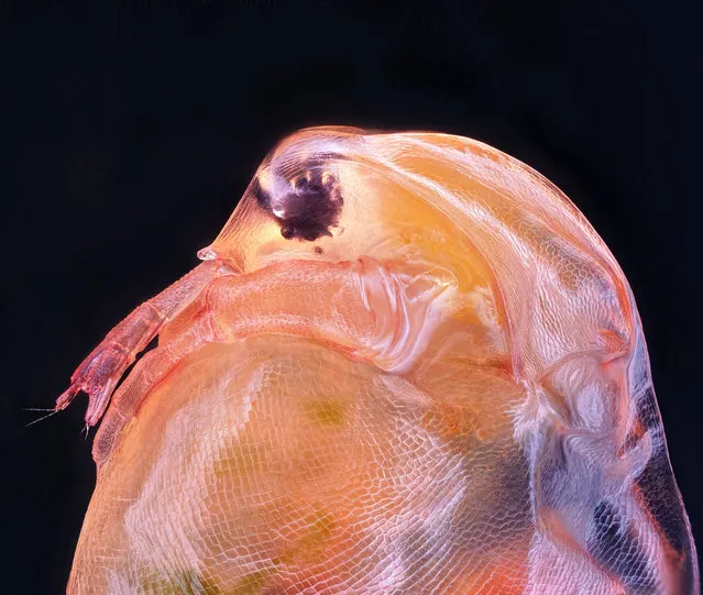
10th Place. Daphnia magna (Phyllopoda). Image Stacking 10X (objective lens magnification). (Photo by Ahmad Fauzan/Nikon Small World Photomicrography 2020)
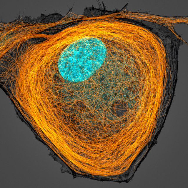
7th Place. Microtubules (orange) inside a cell. Nucleus is shown in cyan. Confocal 63X (objective lens magnification). (Photo by Jason Kirk/Nikon Small World Photomicrography 2020)
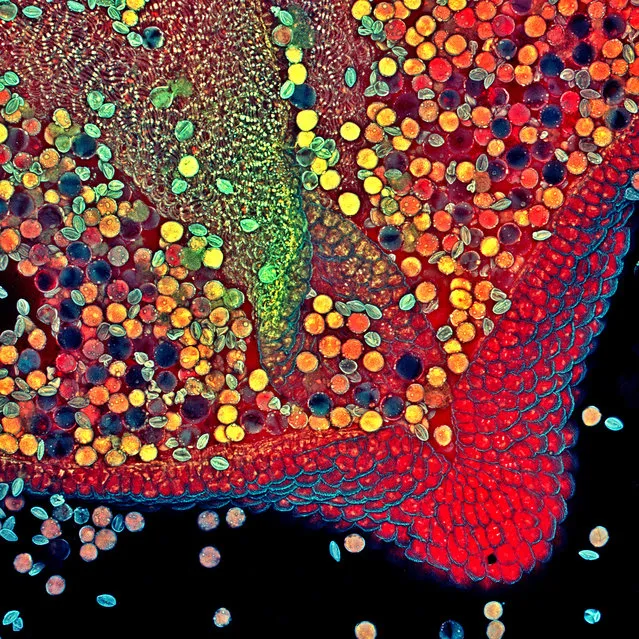
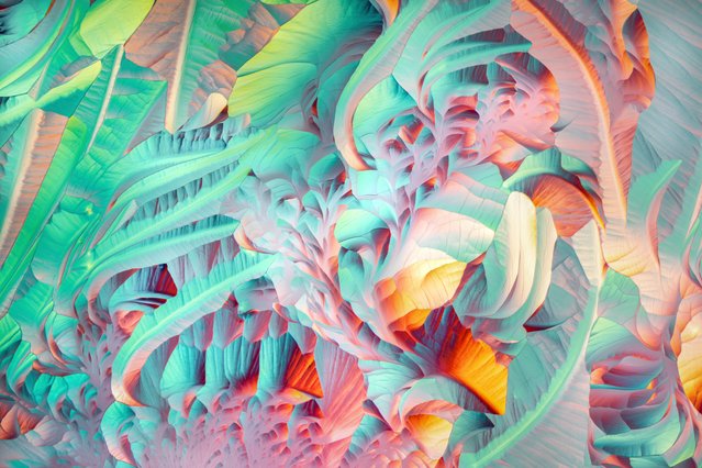
6th Place. Hebe plant anther with pollen. Confocal 10X (objective lens magnification). (Photo by Dr Robert Markus & Zsuzsa Markus/Nikon Small World Photomicrography 2020)
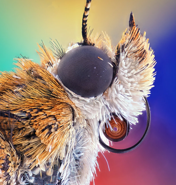
5th Place. Bogong moth. Image Stacking 5X (objective lens magnification). (Photo by Ahmad Fauzan/Nikon Small World Photomicrography 2020)
11th Place. Red algae. Confocal 63X (objective lens magnification). (Photo by Dr Tagide de Carvalho/Nikon Small World Photomicrography 2020)
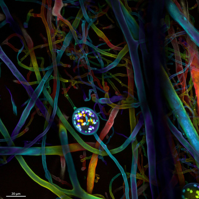
4th Place. Multi-nucleate spores and hyphae of a soil fungus (arbuscular mycorrhizal fungus). Confocal 63X (objective lens magnification). (Photo by Dr Vasileios Kokkoris, Dr Franck Stefani & Dr Nicolas/Nikon Small World Photomicrography 2020)
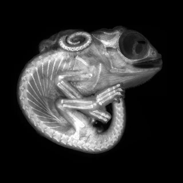
8th Place. Chameleon embryo (autofluorescence). Fluorescence 10X (objective lens magnification). (Photo by Dr Allan Carrillo-Baltodano & David Salamanca/Nikon Small World Photomicrography 2020)
12th Place. Human hair. Image Stacking 20X (objective lens magnification). (Photo by Robert Vierthaler/Nikon Small World Photomicrography 2020)
15 Oct 2020 00:03:00,
post received
0 comments