“Microscope images forge an extraordinary bond between science and art, said Hidenao Tsuchiya, Olympus America's Vice President and General Manager for the Scientific Equipment Group. We founded this competition to focus on the fascinating stories coming out of today's life science research laboratories. The thousands of images that people have shared with the competition over the years reflect some of the most exciting work going on in research today – work that can help shed light on the living universe and ultimately save lives. We look at BioScapes and these beautiful images as sources of education and inspiration to us and the world”. – OlympusBioScapes
Forewing of the green tiger beetle Cicindela campestris by Dr. Jerzy Gubernator of Wroclaw, Poland. (Photo by Olympus BioScapes)
Wheat stigma infected with Claviceps fungus, by Dr. Fernán Federici, University of Cambridge, Plant Sciences Department, Cambridge, UK, and Dr. Anna Gordon, NIAB, Cambridge, UK. (Photo by Olympus BioScapes)
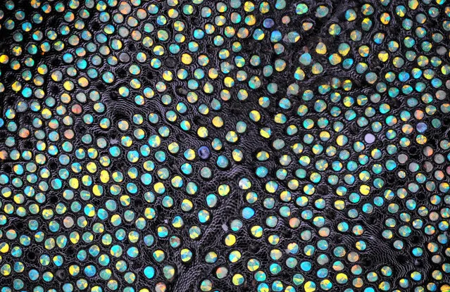
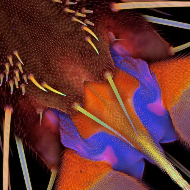
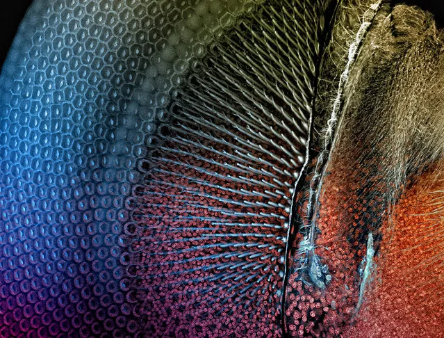
Dried thorax scales of the Weevil Eupholus, by Dr. Douglas Clark, from San Francisco, California. (Photo by Olympus BioScapes)
Purslane (Portulaca) seed, by Yanping Wang from the Beijing Planetarium in Beijing, China. (Photo by Olympus BioScapes)
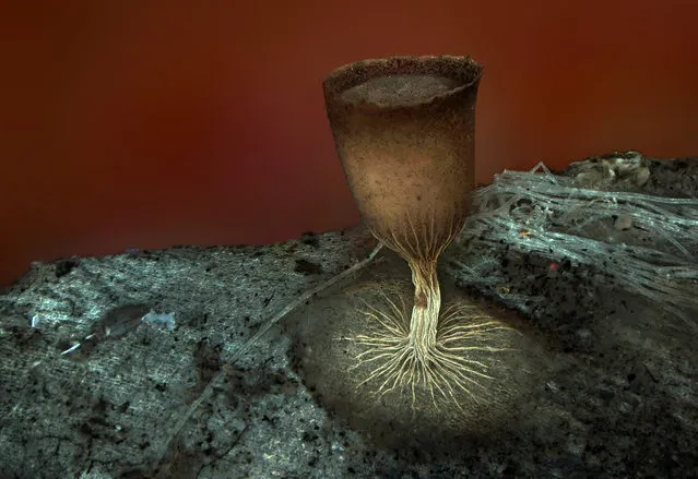
Sporangium of the slime mold Craterium minutum, by Dr. Dalibor Matýsek, Mining University – Technical University of Ostrava, Ostrava, Czech Republic. (Photo by Olympus BioScapes)
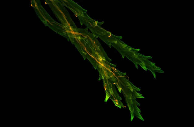
Neuronal culture, fluorescence, six images stitched at 40x magnification, by Jan Schmoranzer, Freie University Berlin, Institute for Chemistry and Biochemistry, Berlin, Germany. (Photo by Olympus BioScapes)
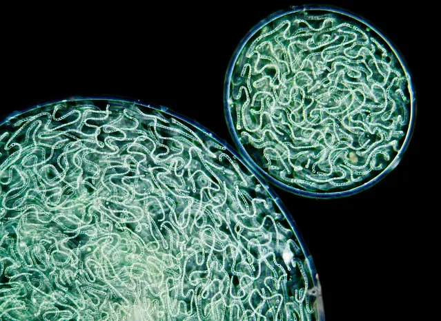
Tenth prize: Spherical colonies of Nostoc commune, a bluegreen alga, by Gerd Guenther, Duesseldorf, Germany. (Photo by Olympus BioScapes)
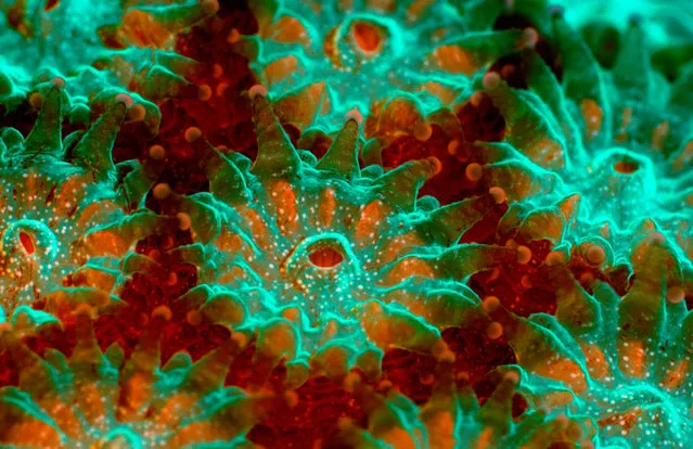
Underwater image of live coral Montastraea annularis. Note polyp tissue (green) around the mouth and base of the tentacles and zooxanthellae (red fluorescence from chlorophyll) in the tissue between polyps. James Nicholson, NOAA/NOS/NCCOS Center for Coastal Environmental Health & Biomolecular Research, Fort Johnson Marine Lab, Charleston, South Carolina. (Photo by Olympus BioScapes)
Plant seed from freshwater pond near Moscow, by Daniel Stoupin of Moscow, Russia. (Photo by Olympus BioScapes)
Pretarsus of the third leg of a female drone fly (Eristalis tenax), ventral view, by Dr. Jan Michels, Institute of Zoology, Christian-Albrechts-University of Kiel, Germany. (Photo by Olympus BioScapes)
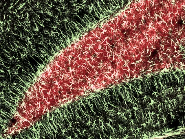
Adult mouse hippocampus, a region of the brain involved in learning and memory. Reactive astroglia (pale yellow) have proliferated and enlarged in response to neuronal activity over time. Dr. Sandra Dieni, Institute of Anatomy and Cell Biology, Albert-Ludwigs University, Freiburg, Germany. (Photo by Olympus BioScapes)
Serum arrested mouse L-1210 cells engaged in spontaneous apoptosis (programmed cell death) after nutrient depletion and acid hydrolysis, by Dr. Frank Abernathy of Jamestown, Ohio. (Photo by Olympus BioScapes)
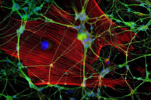
Connective tissue cells co-transduced with five fluorescent proteins, by Dr. Daniela Malide, National Institutes of Health, Bethesda, Maryland. (Photo by Olympus BioScapes)
The eye of a damselfly. The image reveals the regular, crystal-like hexagonal lattice of the eye's elements. Dr. Igor Siwanowicz, Max Planck Institute for Neurobiology, Munich, Germany. (Photo by Olympus BioScapes)
Detail of a pod of the flowering legume Scorpius muricatus (common name “Prickly Caterpillar”), by Viktor Sýkora, from Hyskov, Czech Republic. (Photo by Olympus BioScapes)
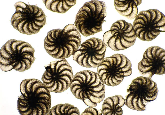
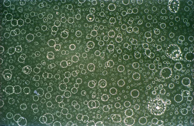
Protozoan Elphidium crispum found growing on the Dorset coast of England, by Michael Gibson of Northampton, UK. (Photo by Olympus BioScapes)
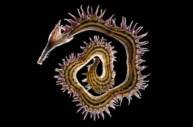
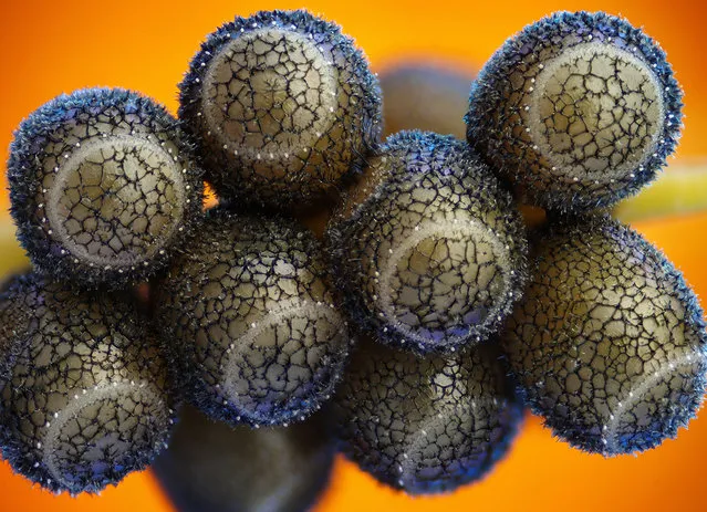
Young sporangia of slime mold Arcyria stipata, by Dr. Dalibor Matýsek, of the Mining University – Technical University of Ostrava, Ostrava, Czech Republic. (Photo by Olympus BioScapes)

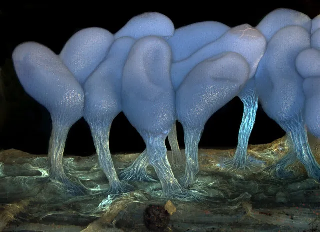
First prize winner: Rotifer Floscularia ringens feeding. Its rapidly beating cilia (hair-like structures) bring water that contains food to the rotifer. The “wheel animacules” were first described by Leeuwenhoek (ca.1702); when their cilia beat, they look like they have two wheels spinning on top. They live in reddish-brown tubes made of spherical “bricks”. Charles Krebs, Issaquah, Washington. (Photo by Olympus BioScapes)
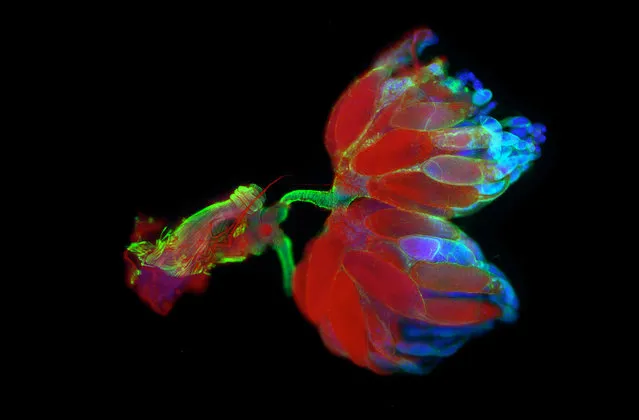
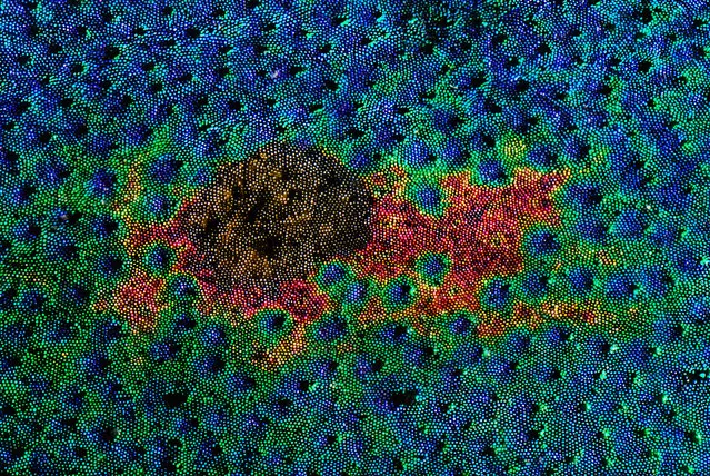
Seventh prize: Fruit fly ovaries and uterus. The muscular and neural structure of the Drosophila melanogaster reproductive system is shown using fluorescence microscopy. The background staining of the eggs in red is a specific function of the mutant fly strain that is pictured here. Gunnar Newquist, University of Nevada, Reno, Nevada. (Photo by Olympus BioScapes)
Sixth prize winner: A cluster of stink bug eggs, by Haris Antonopoulos from Athens, Greece. (Photo by Olympus BioScapes)
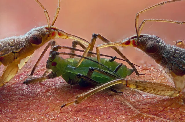
Two damsel bugs (Nabis sp.) seemingly feeding on an aphid by Geir Drange of Asker, Norway. The backdrop is a dried Norway maple leaf. (Photo by Olympus BioScapes)
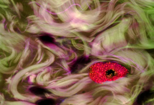
Mast cell in human eye with conjunctivitis. This image shows a single mast cell invading conjunctival tissue in response to an inflammatory agent or pathogen. The mast cell contains vesicles of histamine (red dots). Mast cells are among the first cells of the immune system to react to the presence of an invading pathogen and they facilitate the movement of leukocytes (white blood cells) and other immune cells toward the site of infection. Donald Pottle, The Schepens Eye Research Institute, Boston, Massachusetts. (Photo by Olympus BioScapes)
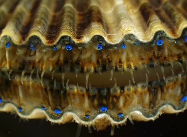
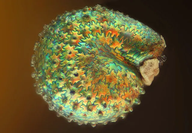
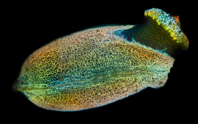
Juvenile live bay scallop Argopecten irradians. The blue spheres are eyes – scallops have up to 100 simple eyes strung around the edges of their mantles. Through research, scientists are trying to help restore scallop populations in Rhode Island. Kathryn Markey, Aquatic Diagnostic Laboratory, Roger Williams University, Bristol, Rhode Island. (Photo by Olympus BioScapes)
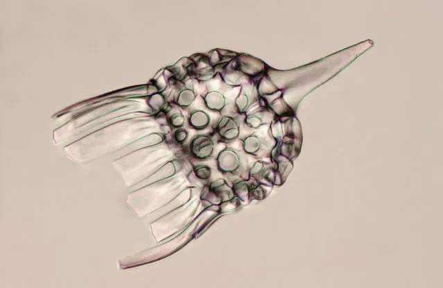
Skeleton of a radiolarian, a single-cell protozoan with an intricate mineral skeleton, by Christopher B. Jackson of Berne, Switzerland. (Photo by Olympus BioScapes)
29 Jul 2012 09:20:00,
post received
0 comments