Now celebrating its 40th year, Nikon Small World is widely regarded as the leading forum to recognize proficiency and photographic excellence of photography taken under the microscope. To select the winners, competition judges analyzed entries from all over the world covering subjects ranging from chemical compounds to up-close-and-personal looks at biological specimens. The 2014 winners will be revealed on October 30th. In 2014, the competition received over 1,200 entries from more than 79 countries around the world.
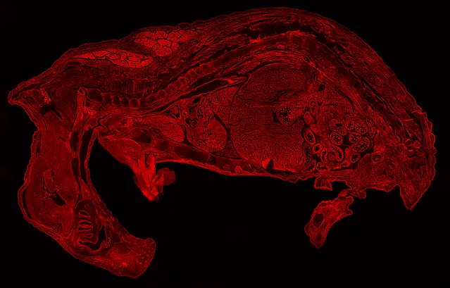
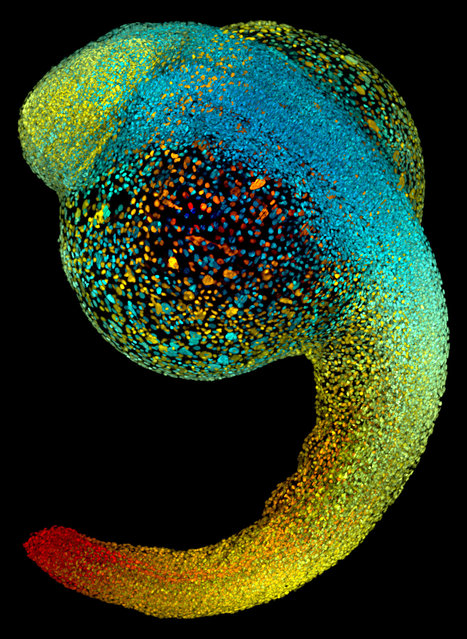
Rat embryo fluorescently labeled with Rhodamine Epi-fluorescence, 10X. Rochester Institute of Technology, Rochester, New York, USA. (Photo by Evan Darling/Nikon Small World 2014)
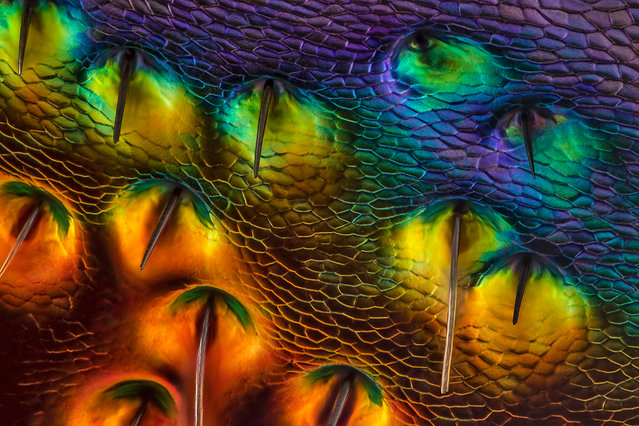
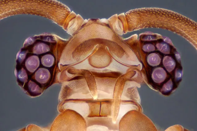
Chrysochroa buqueti (jewel beetle) carapace, near eye Diffused, Reflected Illumination, 450X. Issaquah, Washington, USA. (Photo by Charles Krebs/Nikon Small World 2014)
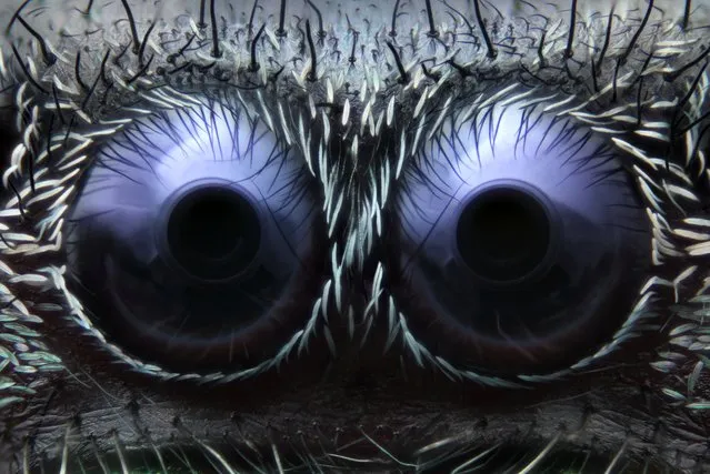
Jumping Spider Eyes; Reflected Light, 20X. Greenwich, Connecticut, USA. (Photo by Noah Fram-Schwartz/Nikon Small World 2014)

Mite in a small forest; Image Stacking, 10X. Biology Department, University of Puerto Rico, Mayaguez Campus; Mayaguez, Puerto Rico. (Photo by José R. Almodóvar/Nikon Small World 2014)
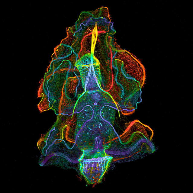
Larval stage of the acorn worm Balanoglossus misakiensis, dorsal view, showing cell borders, muscles and apical eye spots; Confocal, 10X. University of Vienna, Austria. (Photo by Dr. Sabrina Kaul/Nikon Small World 2014)
Live zebrafish embryo at 22 hours post-fertilization; SiMView Light-Sheet Microscopy, 0X. Howard Hughes Medical Institute (HHMI), Ashburn, Virginia, USA. (Photo by Dr. Philipp Keller/Nikon Small World 2014)
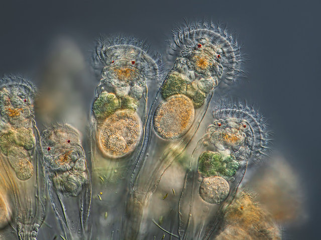
Conochilus unicornis (rotifer), actively feeding. This rotifer species forms a free floating spherical colony; Differential Interference Contrast. Issaquah, Washington, USA. (Photo by Charles Krebs/Nikon Small World 2014)
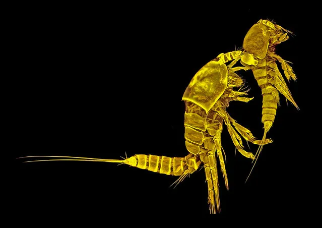
Tigriopus californicus (copepod), couple, lateral view; Confocal Laser Scanning Microscopy, 10X. Senckenberg am Meer, German Centre for Marine Biodiversity Research (DZMB), Wilhelmshaven, Germany. (Photo by Dr. Terue Kihara/Nikon Small World 2014)
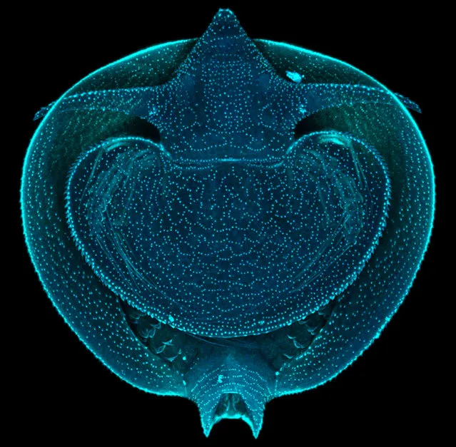
Lynceus brachyurus (clam shrimp), whole mount larva; Confocal, 250X. Department of Integrative Zoology, University of Vienna, Austria. (Photo by Dr. Martin Fritsch/Nikon Small World 2014)
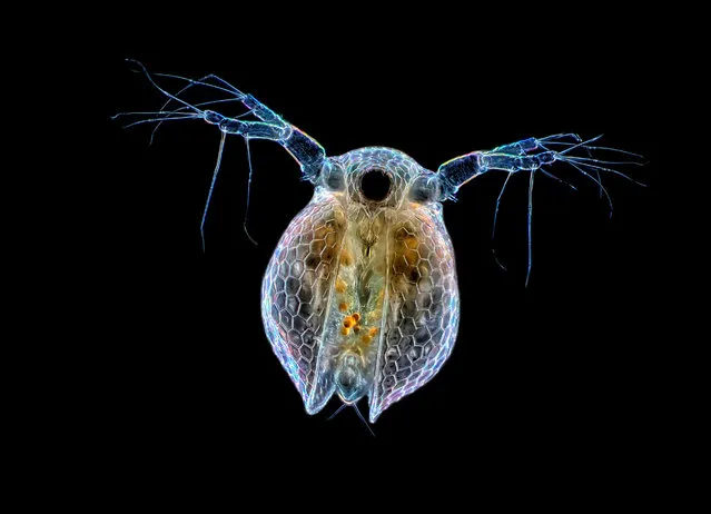
Ceriodaphnia sp. (water flea); Darkfield, 20X. Panama. (Photo by Rogelio Moreno/Nikon Small World 2014)
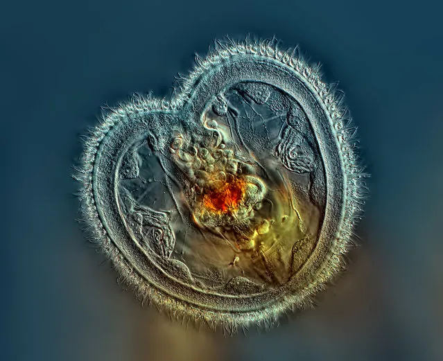
Rotifer showing the mouth interior and heart shaped corona; Differential Interference Contrast, 40X. Panama. (Photo by Rogelio Moreno/Nikon Small World 2014)
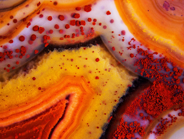
Montana Dryhead agate, unpolished. Axial lighting was provided by Leeds fiberoptic ill, 50X. University Relations and Communications, University of Wisconsin, USA. (Photo by Douglas Moore/Nikon Small World 2014)
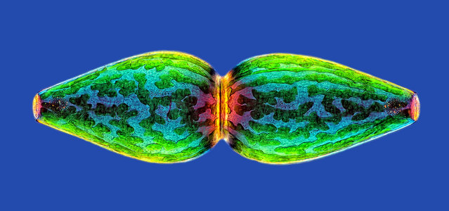
Pleurotaenium ovatum (micro algae); Polarized Light, Lambda Plate, 40X. Panama. (Photo by Rogelio Moreno/Nikon Small World 2014)
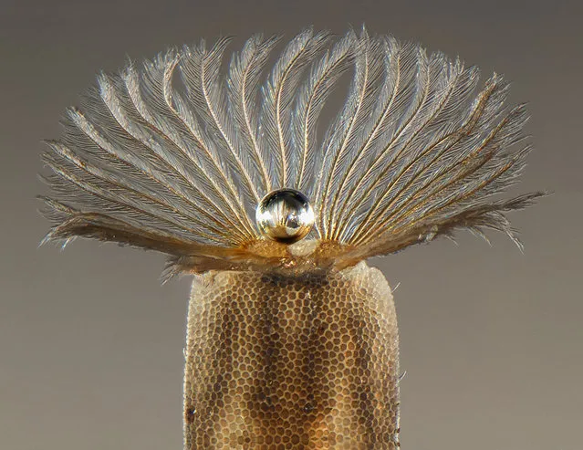
Air pearl in the middle of larva Stratiomyidae resp iratory fringe (Diptera aquatic larva); Stereomicroscopy, 30X. DREAL de Basse-Normandie, Caen, France. (Photo by Fabrice Parais/Nikon Small World 2014)
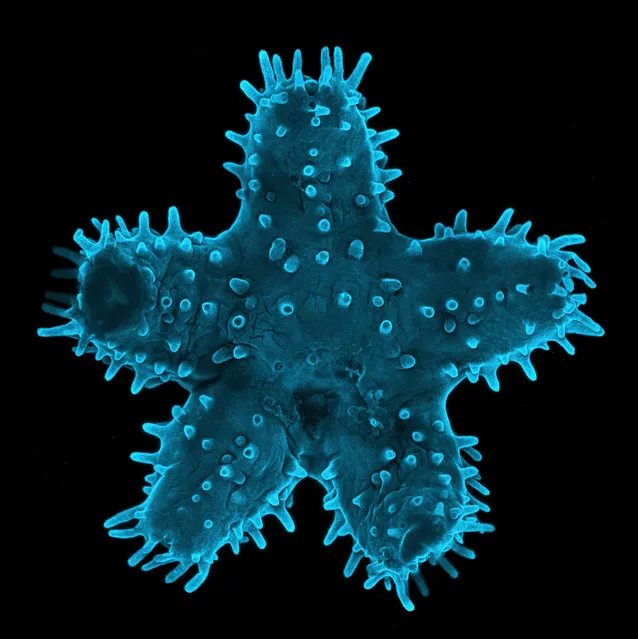
Young Starfish; Confocal, 5X. Confocal Microscopy Lab, Rochester Institute of Technology, Rochester, New York, USA. (Photo by Steven Wilbert/Nikon Small World 2014)
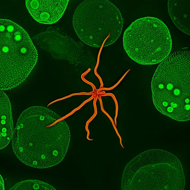
Hydra and Volvox; Confocal, 10X. Confocal Microscopy Lab, Rochester Institute of Technology, Rochester, New York, USA. (Photo by Steven Wilbert/Nikon Small World 2014)
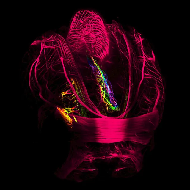
Shipworm Lyrodus pedicellatus, a wood-boring mussel (Mollusca: Bivalvia: Teredinidae). Larval musculature. Confocal laserscanning microscope, 20X. Department of Integrative Zoology, Faculty of Life Sciences, University of Vienna, Austria. (Photo by Andrea Wurzinger-Mayer/Nikon Small World 2014)
Anagallis arvensis (scarlet pimpernel); Macroscopy, 80X. MycoKey, Ebeltoft, Denmark. (Photo by Jens H. Petersen/Nikon Small World 2014)
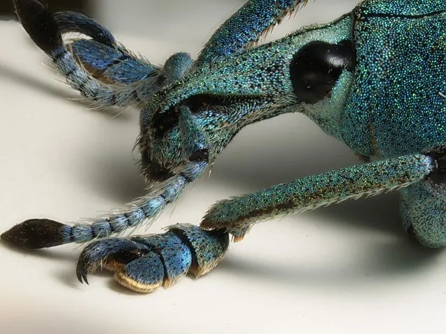
Eupholus sp. (beetle) It's a Coleoptera Curculionidae; Reflected, Diffused Light, 4X. Zoology, Museo Civico di Storia Naturale di Verona, Italy. (Photo by Dr. Luca Toledano/Nikon Small World 2014)
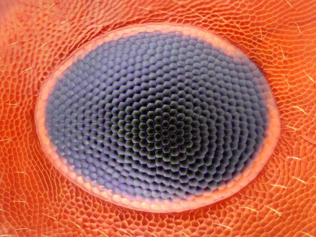
Ant Eye; Reflected Light, 20X. Greenwich, Connecticut, USA. (Photo by Noah Fram-Schwartz/Nikon Small World 2014)
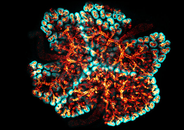
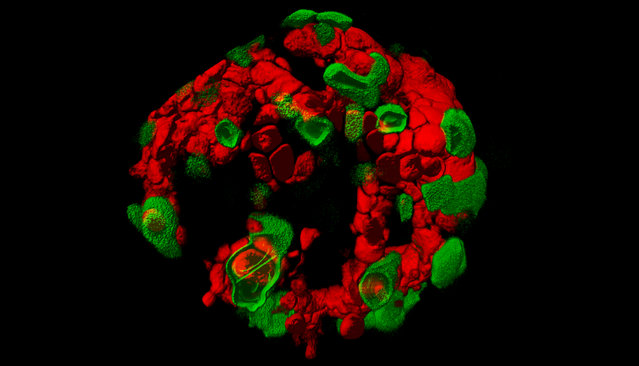
Three transgenic kidneys cultured together, showing colliding branching collecting duct systems; Confocal, 20X. Developmental Biology, The Roslin Institute, Edinburgh, Scotland, UK. (Photo by Dr. Nils Lindstrom/Nikon Small World 2014)
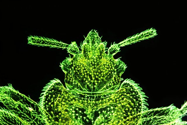
Myrmecolax sp. head, ventral view; Focus Stacking, Reflected Light, 10X. National Museum of Nature and Science, Tsukuba, Ibaraki, Japan. (Photo by Dr. Yuta Nakase/Nikon Small World 2014)
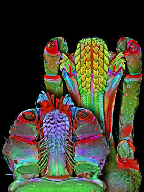
Underside of the Brown dog tick and Lonestar tick mouthparts; Confocal, 100X. Janelia Farm Research Campus, Howard Hughes Medical Institute (HHMI), Ashburn, Virginia, USA. (Photo by Dr. Igor Robert Siwanowicz/Nikon Small World 2014)
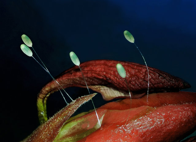
Chrysopa perla (green lacewing) eggs; Stereomicroscope, Focus Stack, 3X. Keszthely, Zala, Hungary. (Photo by Dr. Csaba Pintér/Nikon Small World 2014)
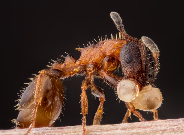
Leptothorax acervorum (ant) carrying its larva; Reflected Light, Focus Stacking, 5X. Asker, Norway. (Photo by Geir Drange/Nikon Small World 2014)
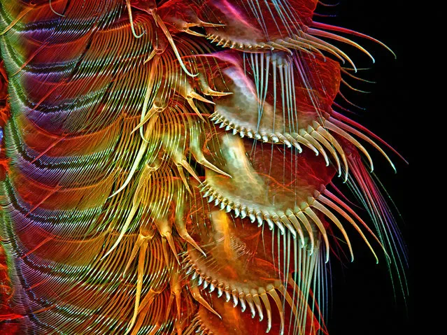
Appendages of a common brine shrimp; Confocal, 100X. Janelia Farm Research Campus, Howard Hughes Medical Institute (HHMI), Ashburn, Virginia, USA. (Photo by Dr. Igor Robert Siwanowicz/Nikon Small World 2014)
Living pancreatic islets; 2-Photon Microscopy, 63X. Edinburgh Super-Resolution Imaging Consortium, Heriot-Watt University, Edinburgh, Scotland. (Photo by Dr. Rory Duncan/Nikon Small World 2014)
Bed bug (Cimex lectularius); Rheinberg illumination (Dark field with interference filter), 50X. Cremona, Italy. (Photo by Stefano Barone/Nikon Small World 2014)
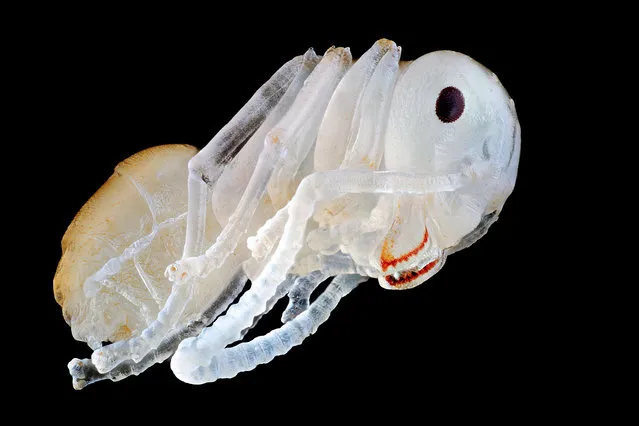
Ant nymph; Image Stacking, Stitching, 10X. Education Nationale Auxonne, Burgundy, France. (Photo by Frederic Labaune/Nikon Small World 2014)
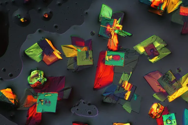
Magnesium chloride and potassium alum mixture; Polarized Light, 25X. National Astronomical Observatories, Chinese Academy of Sciences, Beijing, China. (Photo by Chao Zhang/Nikon Small World 2014)
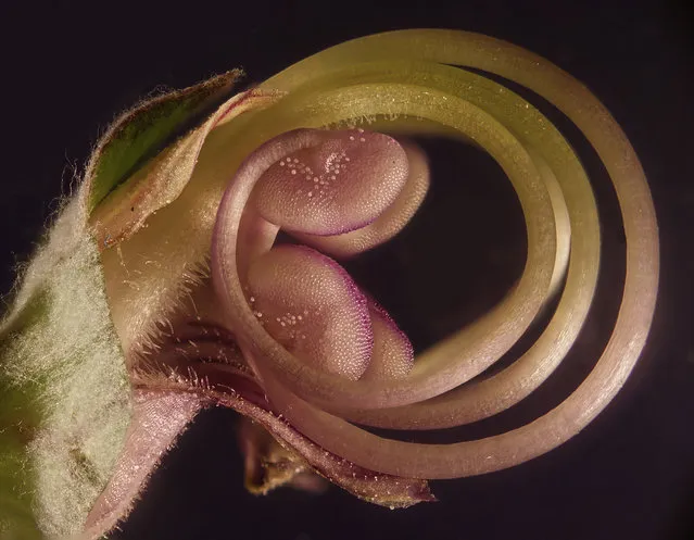
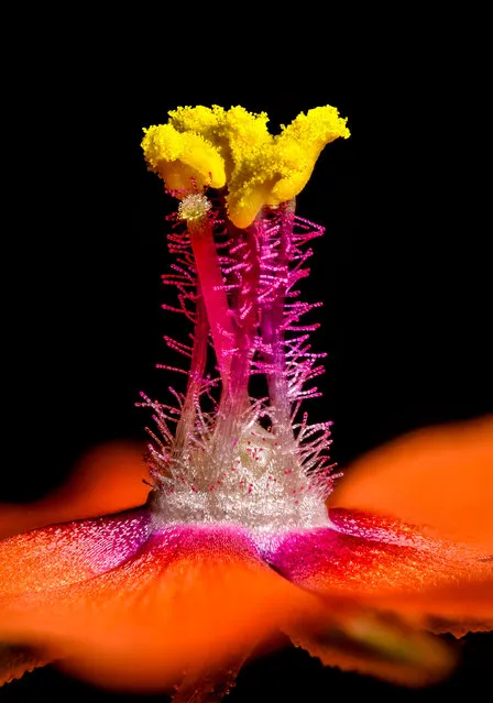
Flower embryo; Fiber Optic Illumination, Reflected Light, 40X. Monsun Yahud, Israel. (Photo by Samuel Silberman/Nikon Small World 2014)
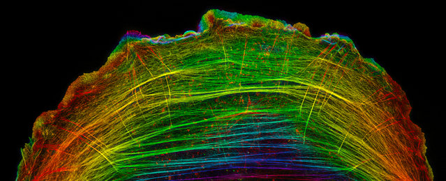
A crawling bone cancer (osteosarcoma) cell showing actin filament bundles in the lamella; Structured Illumination Microscopy, 8000X. Vanderbilt University School of Medicine, Nashville, Tennessee, USA. (Photo by Dr. Dylan T. Burnette/Nikon Small World 2014)
17 Oct 2014 13:10:00,
post received
0 comments