The winning image: trichome (white appendages) and stomata (purple pores) on a southern live oak leaf (60x objective lens magnification). (Photo by Jason Kirk/Nikon Small World Photomicrography 2021)
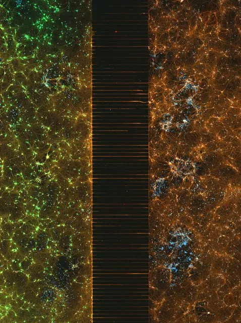
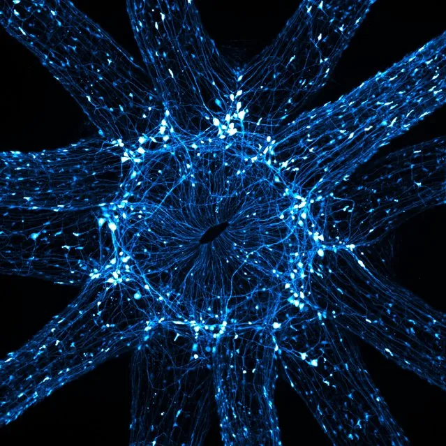
Second place: a microfluidic device containing 300k networking neurons in two isolated populations. Both sides were treated with a unique virus and bridged by axons (fluorescence, 40x objective lens magnification). (Photo by Esmeralda Paric & Holly Stefen/Dementia Research Centre, Macquarie University/Nikon Small World Photomicrography 2021)
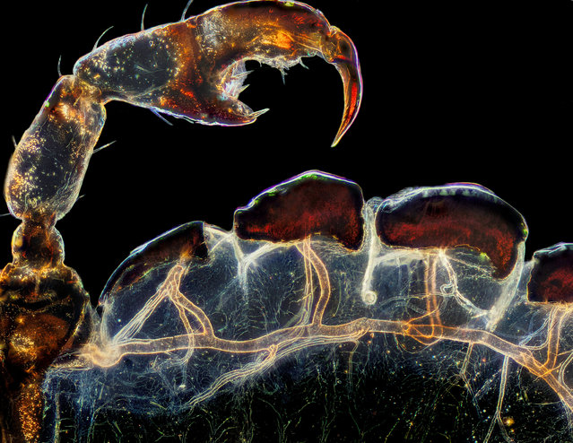
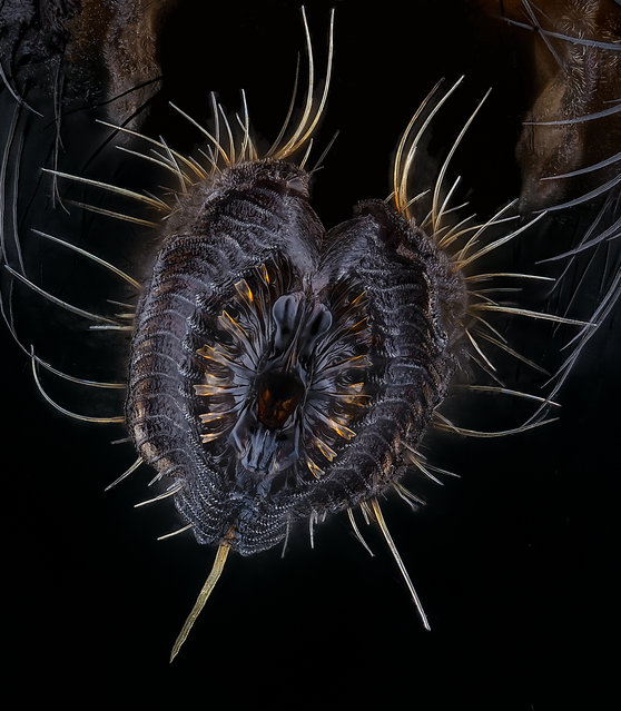
Third place: the rear leg, claw and respiratory trachea of a louse (Haematopinus suis). (Photo by Frank Reiser/Nassau Community College/Nikon Small World Photomicrography 2021)
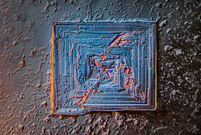
18th place: a table salt crystal (image stacking, darkfield, oblique, Rheinberg, polarised light, 10x objective lens magnification). (Photo by Saulius Gugis/Nikon Small World Photomicrography 2021)
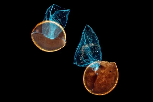
Honourable mention: hatched brine shrimp eggs (darkfield, fluorescence, image stacking). (Photo by Waldo Nell/Nikon Small World Photomicrography 2021)
Fifth place: proboscis of a housefly (Musca domestica) (image stacking, 40x objective lens magnification). (Photo by Oliver Dum/Nikon Small World Photomicrography 2021)
Honourable Mention: a midge (Chironomidae diptera). (Photo by Dr. Erick Francisco Mesén/Nikon Small World Photomicrography 2021)
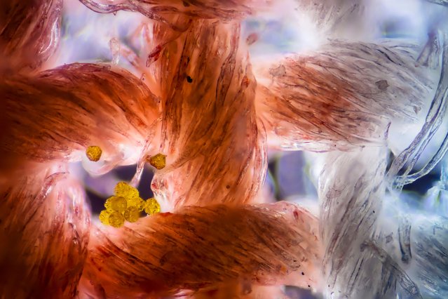
13th place: cotton fabric with pollen grains. (Photo by Felice Placenti/Nikon Small World Photomicrography 2021)
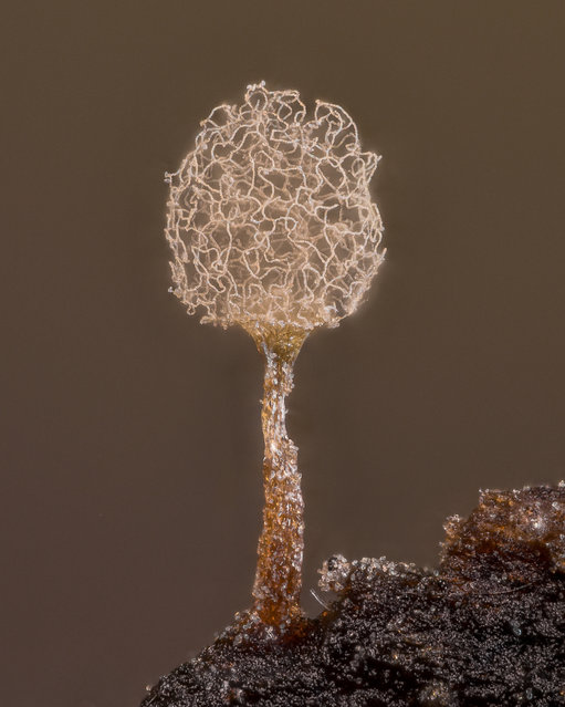
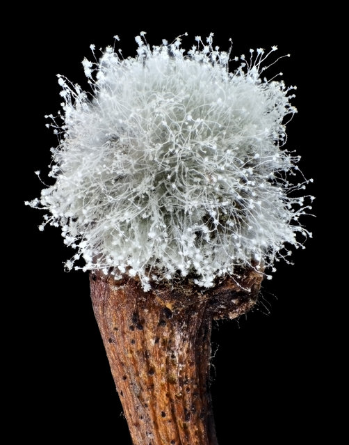
20th place: slime mould (Arcyria pomiformis). (Photo by Alison K. Pollack/Nikon Small World Photomicrography 2021)
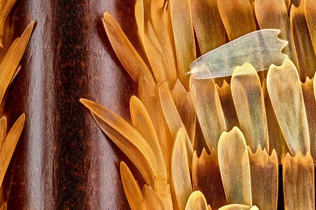
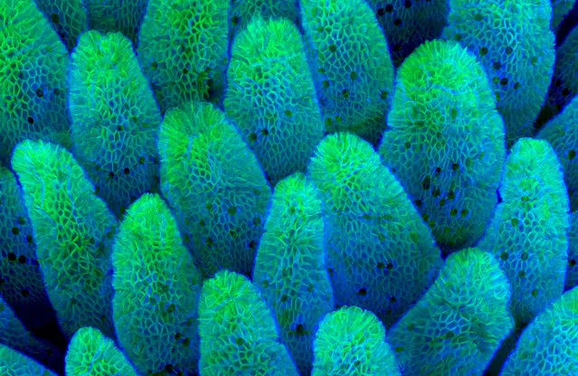
10th place: vein and scales on a butterfly wing (Morpho didius) (20x objective lens magnification). (Photo by Sébastien Malo/Nikon Small World Photomicrography 2021)
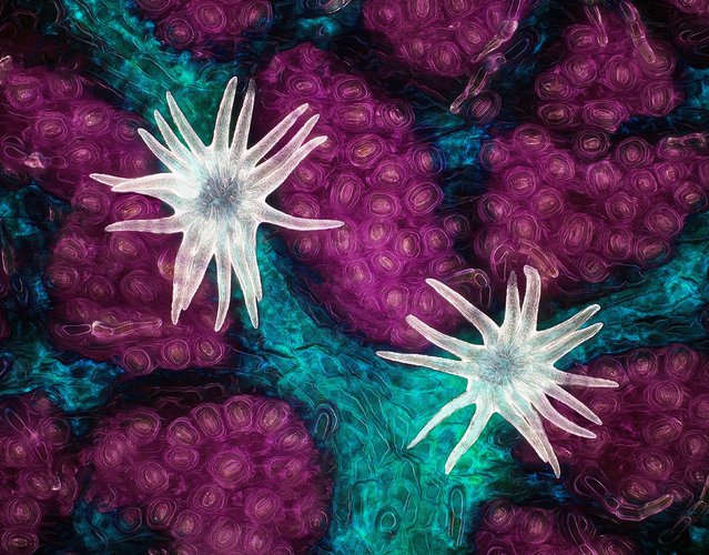
16th place: an in-vivo snapshot of the neurons surrounding the mouth and tentacles of a juvenile starlet sea anemone (Nematostella vectensis) (20x objective lens magnification). (Photo by Ruohan Zhong/Stowers Institute for Medical Research/Nikon Small World Photomicrography 2021)
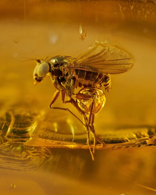
Honourable mention: an amber series project displays tiny insects no more than 3mm long that have been encapsulated within hardened tree sap for 45 million years. (Photo by Levon Biss Photography Ltd./Nikon Small World Photomicrography 2021)
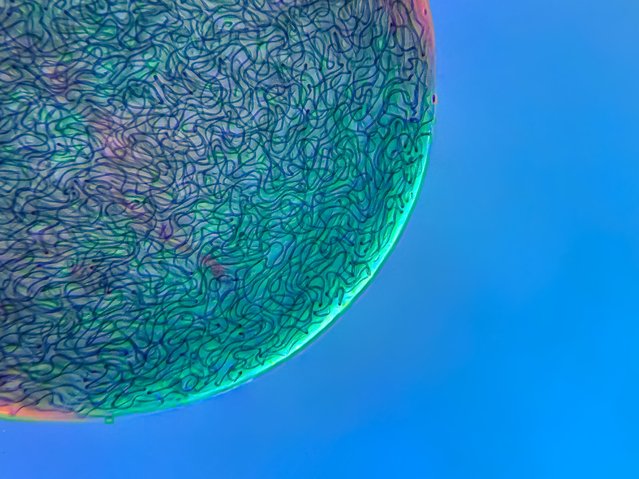
17th place: filamentous strands of Nostoc cyanobacteria captured inside a gelatinous matrix. (4x objective lens magnification). (Photo by Martin Kaae Kristiansen/My Microscopic World/Nikon Small World Photomicrography 2021)
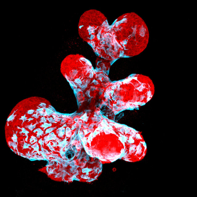
12th place: a breast organoid showing contractile myoepithelial cells (blue) crawling on secretory breast cells (red). (Photo by Jakub Sumbal/Masaryk University/Nikon Small World Photomicrography 2021)
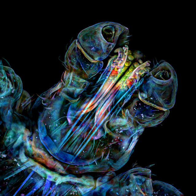
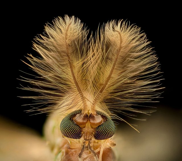
Seventh place: the head of a tick. (Photo by Dr. Tong Zhang & Dr. Paul Stoodley/The Ohio State University/Nikon Small World Photomicrography 2021)
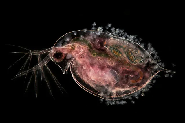
Ninth place: a water flea carrying embryos and peritrichs. (Photo by Jan van IJken Photography and Film/Nikon Small World Photomicrography 2021)
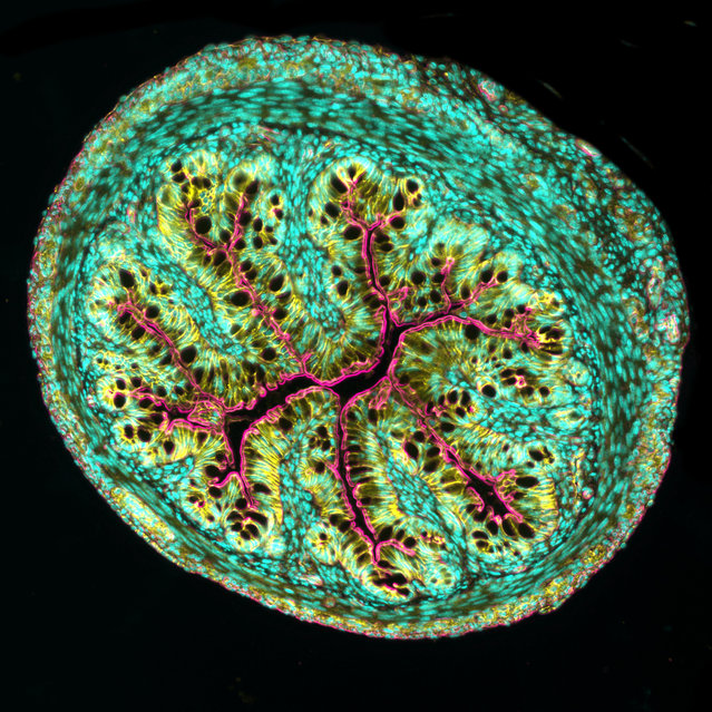
Eighth place: a cross-section of mouse intestine. (Photo by Dr. Amy Engevik/Medical University of South Carolina/Nikon Small World Photomicrography 2021)
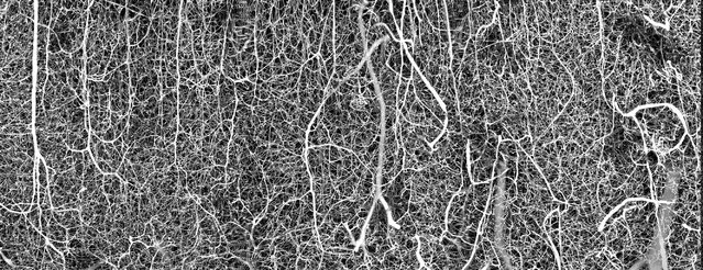
Sixth place: a 3D vasculature of an adult mouse brain (somatosensory cortex). (Photo by Dr. Andrea Tedeschi/The Ohio State University/Wexner Medical Center/Nikon Small World Photomicrography 2021)
Honourable mention: mould on top of a cherry stem (image stacking, reflected light, 2x objective lens magnification). (Photo by Sergii Dymchenko/SDym Photography/Nikon Small World Photomicrography 2021)

Honourable mention: a water flea swimming near a reed stalk. (Photo by Álmos Becz/Eötvös Loránd University/Nikon Small World Photomicrography 2021)
Honourable mention: epithelial cells covering the intestine villi (confocal, fluorescence. 63x objective lens magnification). (Photo by Caleb Dawson/The Walter and Eliza Hall Institute of Medical Research/Nikon Small World Photomicrography 2021)
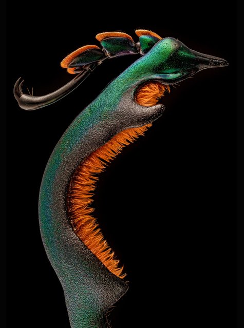
Honourable mention: the hind leg of a male frog-legged beetle (Sagra buqueti). (Photo by Dr Andrew Mark Posselt/University of California, San Francisco/Nikon Small World Photomicrography 2021)
15 Sep 2021 02:36:00,
post received
0 comments