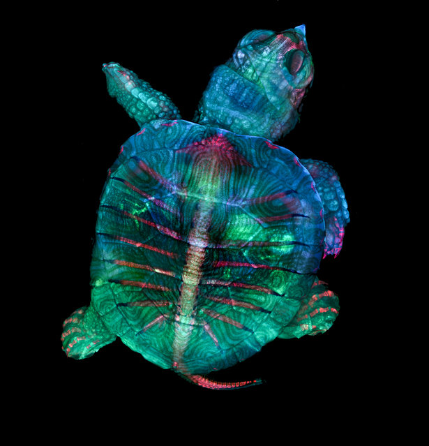
1st Place: Teresa Zgoda & Teresa Kugler, Campbell Hall, New York, USA. Fluorescent turtle embryo. Stereomicroscopy, Fluorescence, 5x (Objective Lens Magnification). (Photo by Teresa Zgoda/Nikon's Small World 2019)
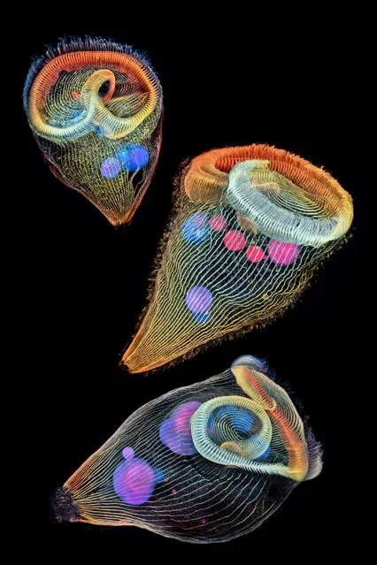
2nd Place: Dr. Igor Siwanowicz, Howard Hughes Medical Institute (HHMI), Janelia Research Campus, Ashburn, Virginia, USA. Depth-color coded projections of three stentors (single-cell freshwater protozoans). Confocal, 40x (Objective Lens Magnification). (Photo by Igor Siwanowicz/Nikon's Small World 2019)
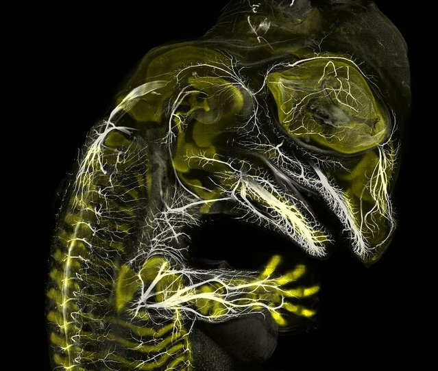
3rd Place: Daniel Smith Paredes & Dr. Bhart-Anjan S. Bhullar, Yale University, Department of Geology and Geophysics, New Haven, Connecticut, USA. Alligator embryo developing nerves and skeleton. Immunofluorescence, 10x (Objective Lens Magnification). (Photo by Daniel Smith Paredes/Nikon's Small World 2019)
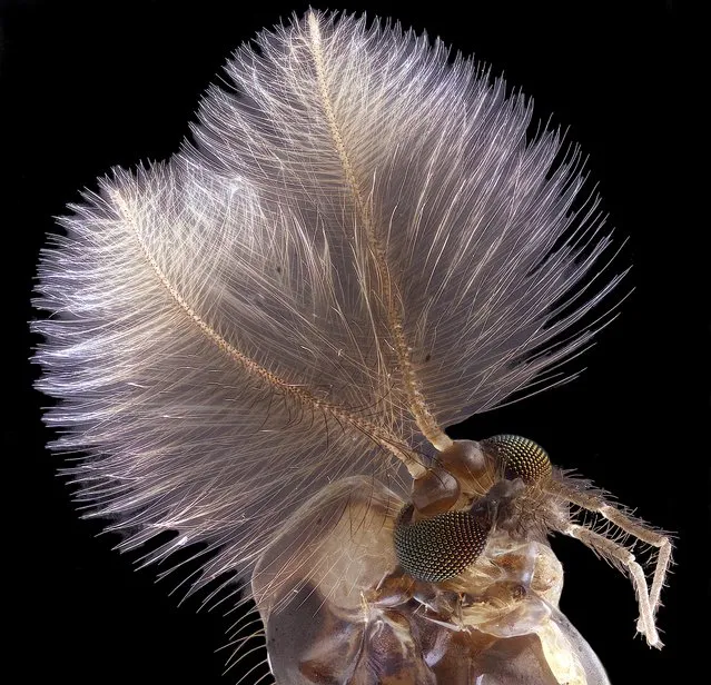
4th Place: Jan Rosenboom, Universität Rostock, Rostock, Mecklenburg Vorpommern, Germany. Male mosquito. Focus Stacking, 6.3x (Objective Lens Magnification). (Photo by Jan Rosenboom/Nikon's Small World 2019)
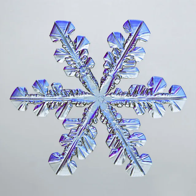
5th Place: Caleb Foster, Caleb Foster Photography, Jericho, Vermont, USA. Snowflake. Transmitted Light, 4x (Objective Lens Magnification). (Photo by Caleb Foster/Nikon's Small World 2019)
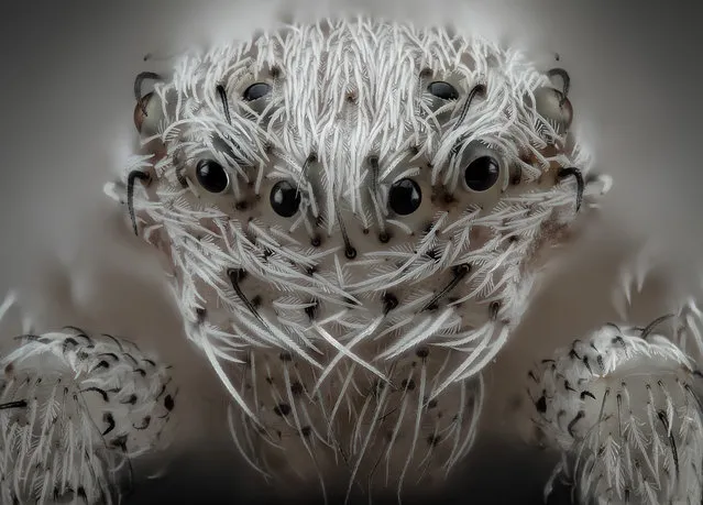
6th Place: Javier Rupérez, Almáchar, Málaga, Spain. Small white hair spider. Reflected Light, Image Stacking, 20x (Objective Lens Magnification). (Photo by Javier Rupérez/Nikon's Small World 2019)
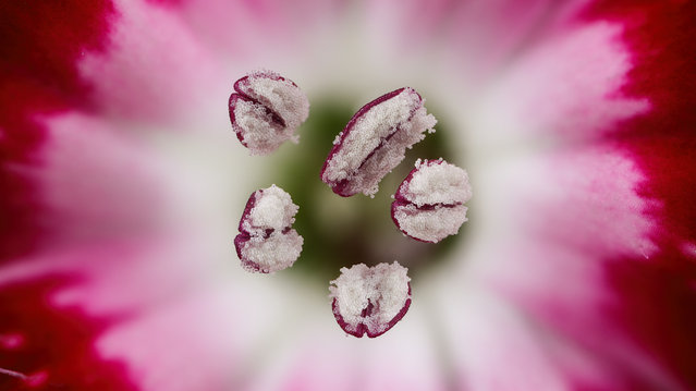
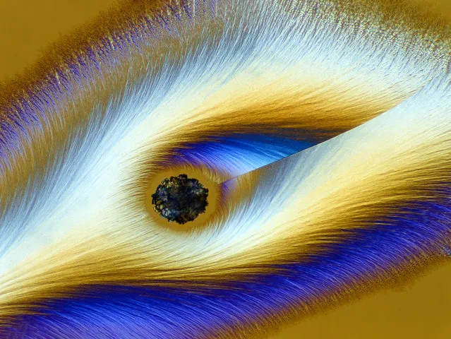
7th Place: Dr. Guillermo López, Alicante, Spain. Chinese red carnation stamen. Focus Stacking, 3x (Objective Lens Magnification). (Photo by Guillermo López/Nikon's Small World 2019)
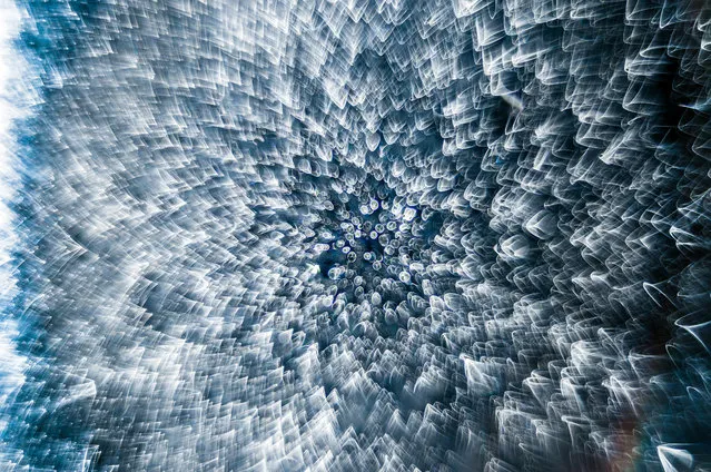
8th Place: Garzon Christian, Quintin, Cotes-d’Armor, France. Frozen water droplet. Incident Light, 8x (Objective Lens Magnification). (Photo by Garzon Christian/Nikon's Small World 2019)
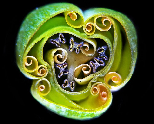
9th Place: Andrei Savitsky, Cherkassy, Ukraine. Tulip bud cross section. Reflected Light, 1x (Objective Lens Magnification). (Photo by Andrei Savitsky/Nikon's Small World 2019)
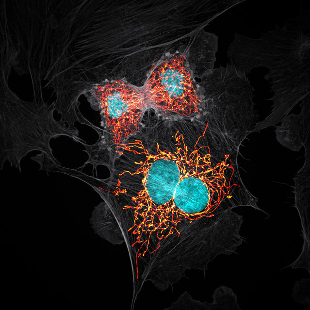
10th Place: Jason M. Kirk, Baylor College of Medicine, Optical Imaging & Vital Microscopy Core, Houston, Texas, USA. BPAE cells in telophase stage of mitosis. Confocal with Enhanced Resolution, 63x (Objective Lens Magnification). (Photo by Jason M. Kirk/Nikon's Small World 2019)
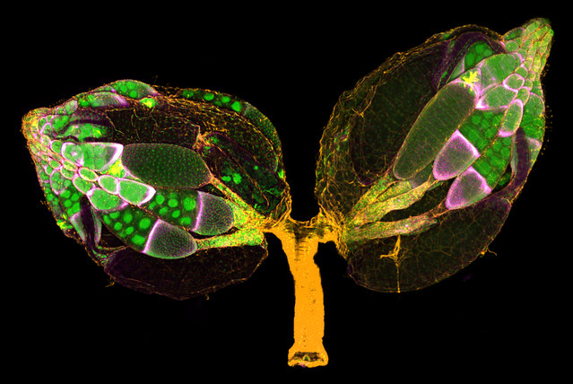
11th Place: Dr. Yujun Chen & Dr. Jocelyn McDonald, Kansas State University, Department of Biology, Manhattan, Kansas, USA. A pair of ovaries from an adult Drosophila female stained for F-actin (yellow) and nuclei (green); follicle cells are marked by GFP (magenta). Confocal, 10x (Objective Lens Magnification). (Photo by Yujun Chen/Nikon's Small World 2019)
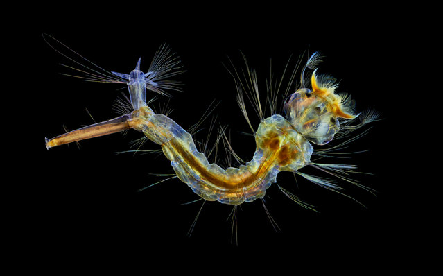
12th Place: Anne Algar, Hounslow, Middlesex, United Kingdom. Mosquito larva. Darkfield, Polarizing Light, Image Stacking, 4x (Objective Lens Magnification). (Photo by Anne Algar/Nikon's Small World 2019)
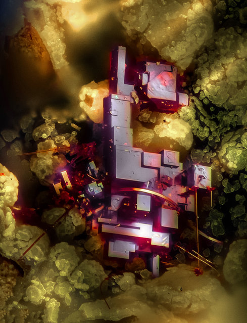
13th Place: Dr. Emilio Carabajal Márquez, Madrid, Spain. Cuprite (mineral composed of copper oxide). Focus Stacking, 20x (Objective Lens Magnification). (Photo by Emilio Carabajal Márquez/Nikon's Small World 2019)
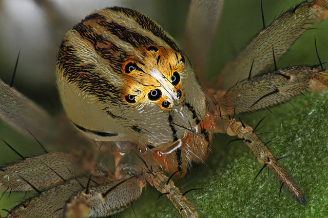
14th Place: Antoine Franck, CIRAD – Agricultural Research for Development, Saint Pierre, Réunion. Female Oxyopes dumonti (lynx) spider. Focus Stacking, 1x (Objective Lens Magnification). (Photo by Antoine Franck/Nikon's Small World 2019)
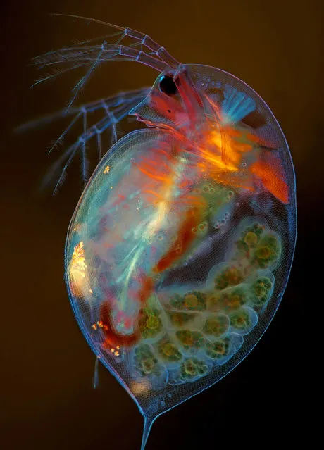
15th Place: Marek Miś, Marek Miś Photography, Suwalki, Podlaskie, Poland. Pregnant Daphnia magna (small planktonic crustacean). Modified Darkfield, Polarized Light, Image Stacking, 4x (Objective Lens Magnification). (Photo by Marek Miś/Nikon's Small World 2019)
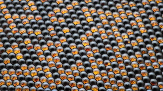
16th Place: Dr. Razvan Cornel Constantin, Bucharest, Romania. Housefly compound eye pattern. Focus Stacking, Reflected Light, 50x (Objective Lens Magnification). (Photo by Razvan Cornel Constantin/Nikon's Small World 2019)
17th Place: Karl Deckart, Eckental, Bavaria, Germany. Vitamin C. Brightfield, Polarized Light, 4x (Objective Lens Magnification). (Photo by Karl Deckart/Nikon's Small World 2019)
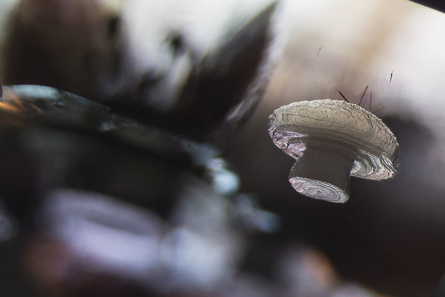
18th Place: E. Billie Hughes, Lotus Gemology, Bangkok, Thailand. Cristobalite crystal suspended in its quartz mineral host. Darkfield, 40x (Objective Lens Magnification). (Photo by E. Billie Hughes/Nikon's Small World 2019)
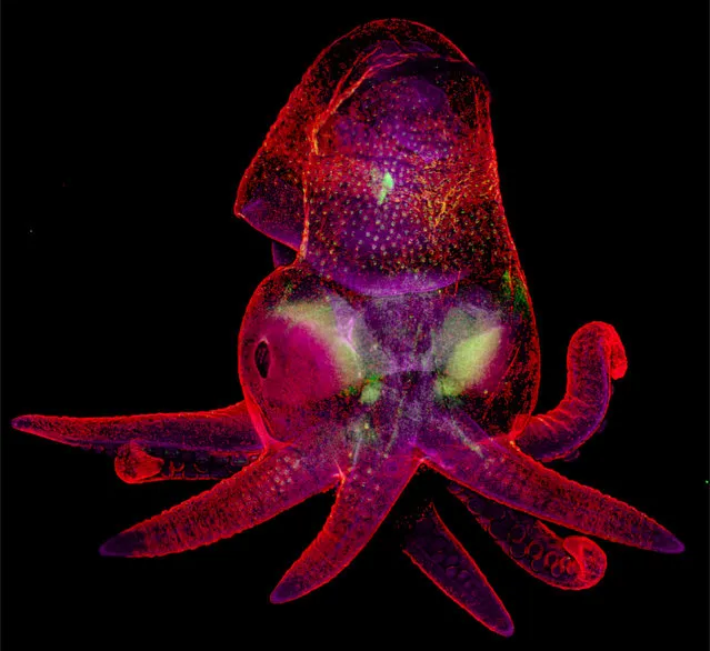
19th Place: Martyna Lukoseviciute & Dr. Carrie Albertin, University of Oxford, Weatherall Institute of Molecular Medicine, Oxford, Oxfordshire, United Kingdom. Octopus bimaculoides embryo. Confocal, Image Stitching, 5x (Objective Lens Magnification). (Photo by Martyna Lukoseviciute/Nikon's Small World 2019)
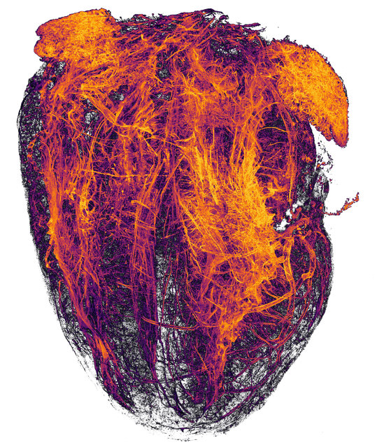
20th Place: Simon Merz, Lea Bornemann & Sebastian Korste, University Hospital Essen, Institute for Experimental Immunology & Imaging, Essen, Nordrhein-Westfalen, Germany. Blood vessels of a murine (mouse) heart following myocardial infarction (heart attack). Tissue Clearing, Light Sheet Fluorescence Microscopy, 2x (Objective Lens Magnification). (Photo by Simon Merz/Nikon's Small World 2019)
23 Oct 2019 00:03:00,
post received
0 comments