
Cat skin and blood supply. Whiskers, unlike normal hair, are touch receptors, each containing a sensory organ called a proprioceptor. Scientists injected blood vessels with a red dye called carmine dye (here appearing black) in order to visualise the capillaries in the tissue, a newly developed technique at the time. The picture is a composite made up of 44 individual images which were stitched together. Here, fine hairs (yellow), thicker whisker (yellow) and blood vessels (black) are all visible. (Photo by David Linstead/Wellcome Images)
A developing spinal cord of a baby mouse. (Photo by Gabriel Galea/Wellcome Images/UCL)
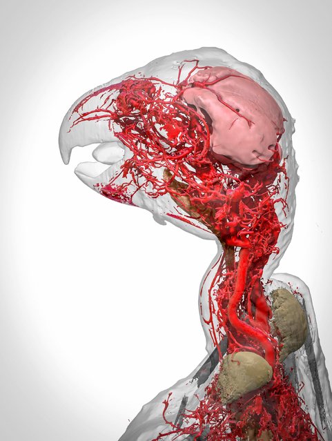
Blood vessels of an African Grey parrot. The 3D reconstruction of an African grey parrot just after it died shows the intricate system of blood vessels in its head and neck. This was only possible thanks to a special chemical called BriteVu which lets researchers study veins in incredible detail. (Photo by Scott Birch/Scott Echols/Wellcome Images)
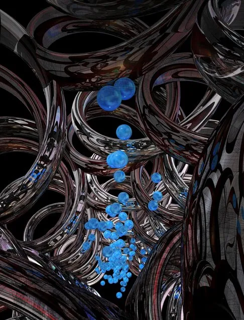
Synthetic DNA channel transporting cargo across membranes. Every cell is surrounded by a membrane, which serves to protect the cell’s contents from its external environment, provide support, and connect the cell to others to form tissues and organs. Tube-like channels made of proteins span this membrane and control two-way communication between the cell and its environment. Researchers are using DNA as a building material to make synthetic channels that behave in exactly the same way. This image is an artist’s representation of what these channels look like. (Photo by Michael Northrop/Wellcome Images)
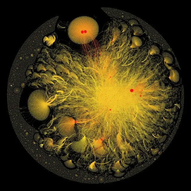
Twitter feed. This is a graphical visualisation of data extracted from tweets containing the hashtag #breastcancer. (Photo by PA Wire/Wellcome Images)
A 3D model of a healthy mini-pig eye. The dent on the right-hand side of this 3D model of a pig eye is the pupil, the opening that allows light into the eye. Blood vessels are pumped up, bringing energy and food to the muscles surrounding the iris, which controls the amount of light entering the eye. The smallest vessels seen here are just 20-30 micrometres (0.02-0.03 mm) in diameter. The other large vessels are feeder vessels for the retina, the light-sensing region at the back of the eye. (Photo by Dr Peter M. Maloca/Wellcome Images)
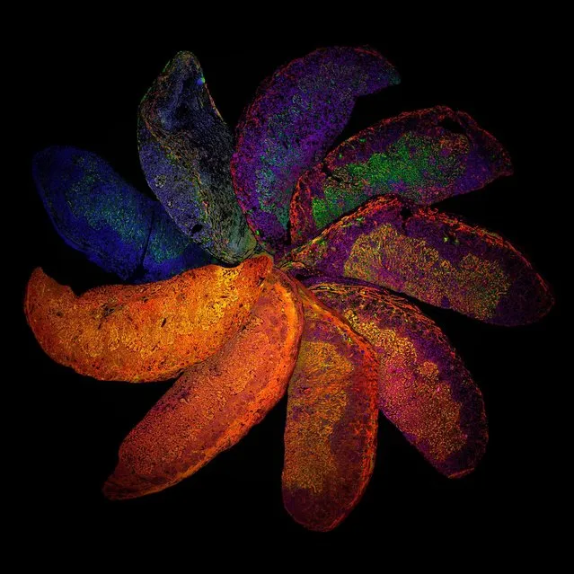
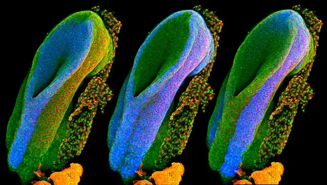
This Terry’s Chocolate Orange-type display is actually a variety of mice placentas, dubbed The Placenta Rainbow. Despite being the same age (Around the 12-week pregnancy mark) the placentas look different. That’s because they show the difference mother’s immune systems can have on placental development. Blue represents the nucleus, where DNA is stored and controlled; blood vessels are stained in red; and trophoblasts, the first cells to form in the developing embryo, are stained in green. Additional colours are present due to an expression of two or more of these proteins in the same cell. The range of colours indicates the significant effects that differences in a mother’s immune system can have on placental development. Such techniques could help us understand and identify ways to treat complications that arise during human pregnancies. (Photo by PA Wire/Wellcome Images)
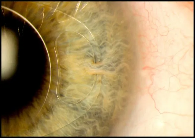
It’s of an “iris clip” which is often known as an artificial intraocular lens. The clip is a small thin lens made from silicone to treat nearsightedness and cataracts. This particular patient, a 70-year-old man, regained almost full vision following his surgery. (Photo by Mark Bartley/Cambridge University Hospitals NHS Foundation Trust/Wellcome Images)
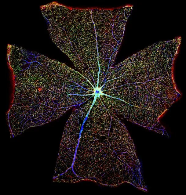
Mouse eyeball. This picture was created by digitally stitching together over 400 images to form one large image, so as to show the entire surface of a mouse retina. (Photo by Gabriel Luna/Wellcome Images)
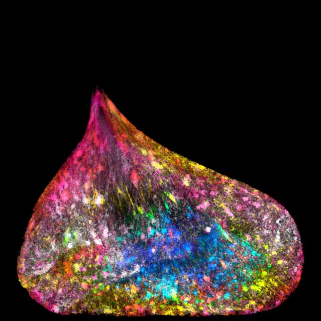
Ezequiel Miron captured a multicoloured portrait of DNA in a human lung cell. The DNA in the cell has somehow become caught, and is being pulled between the two cells, making it appear like a teardrop. This has caused the DNA to unfold inside the nucleus, and DNA fibres can be seen running through it. As the new cells have moved apart, the tension distributed by the rope-like DNA has deformed the nucleus’s usually circular shape. (Photo by Ezequiel Miron/Wellcome Images)
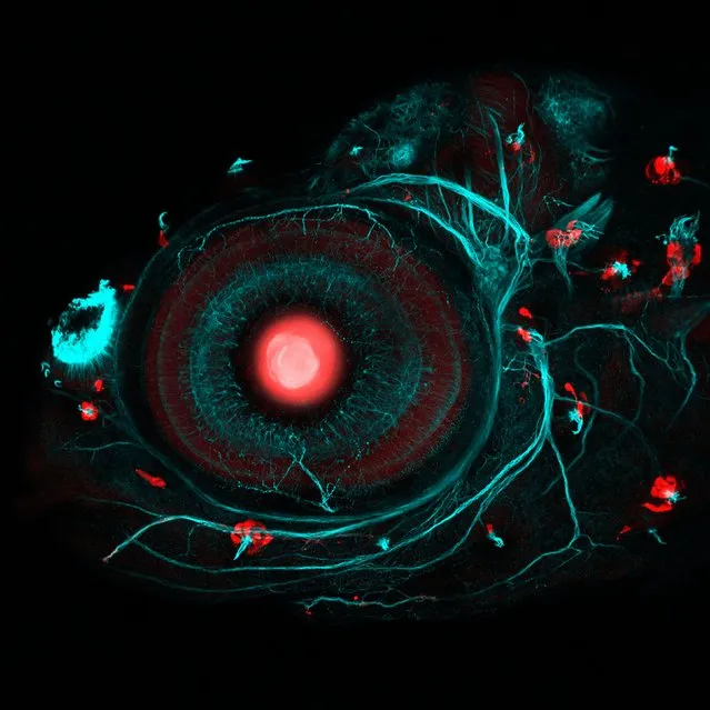
This picture shows a Zebrafish eye and neuromasts. This four-day-old zebrafish embryo has been modified so scientists can understand the genetics behind the development of the eye. The Zebrafish eye is particularly interesting because it responds to movement in the water and is essential for avoiding predators and help it keep up with its school. The blueish-green lines are its nervous system, which is also being studied by experts at University College London. (Photo by Ingrid Lekk/Steve Wilson/Wellcome Images/UCL)
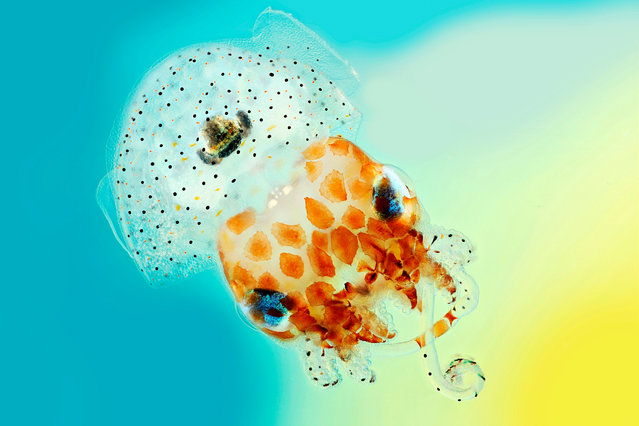
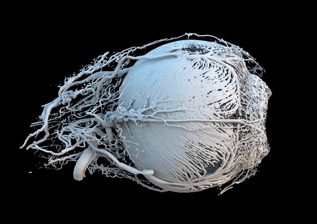
With its huge eyes, comical name and diminutive size, Mark R. Smith’s image of a baby Hawaiian bobtail squid can’t help but raise a smile. A curiously endearing creature, the cephalopod is just 1.5cm across, its mantle cavity bearing more than a passing resemblance to a rather natty shower cap. But it is also a beautiful example of symbiosis – nature’s version of “I’ll scratch your back if you scratch mine” – for on the underside of the squid is a light organ which houses bioluminescent bacteria. The squid offers the bacteria protection and food, while the bacteria emit a glow – a handy trait that the squid uses to offset its silhouette, helping it to evade predators in the depths below. Mark R. Smith’s entry combines several images of a Hawaiian bobtail squid with different focus lengths to create a final picture with greater depth of field than normal. (Photo by Mark R. Smith/Wellcome Images/Macroscopic Solutions)
08 Mar 2017 00:05:00,
post received
0 comments