Loading...
Done
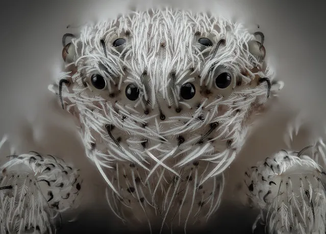
6th Place: Javier Rupérez, Almáchar, Málaga, Spain. Small white hair spider. Reflected Light, Image Stacking, 20x (Objective Lens Magnification). (Photo by Javier Rupérez/Nikon's Small World 2019)

6th Place: Javier Rupérez, Almáchar, Málaga, Spain. Small white hair spider. Reflected Light, Image Stacking, 20x (Objective Lens Magnification). (Photo by Javier Rupérez/Nikon's Small World 2019)
Details
23 Oct 2019 00:03:00
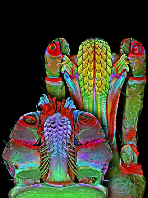
Underside of the Brown dog tick and Lonestar tick mouthparts; Confocal, 100X. Janelia Farm Research Campus, Howard Hughes Medical Institute (HHMI), Ashburn, Virginia, USA. (Photo by Dr. Igor Robert Siwanowicz/Nikon Small World 2014)

Now celebrating its 40th year, Nikon Small World is widely regarded as the leading forum to recognize proficiency and photographic excellence of photography taken under the microscope. To select the winners, competition judges analyzed entries from all over the world covering subjects ranging from chemical compounds to up-close-and-personal looks at biological specimens. The 2014 winners will be revealed on October 30th. In 2014, the competition received over 1,200 entries from more than 79 countries around the world. (Photo by Dr. Igor Robert Siwanowicz/Nikon Small World 2014)
Details
17 Oct 2014 13:10:00
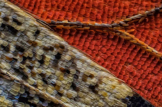
11th Place: Scales of a butterfly wing underside (Vanessa atalanta). (Photo by Francis Sneyers/Nikon's Small World 2016)

Each year Nikon Small World recognizes the excellence of photography taken under the microscope. The contest showcases the beauty and complexity of life. Anyone interested in microscopy and photography can enter the contest and in its 42nd year, Nikon Small World received 2,000 entries from 70 countries. Here: 11th Place; Scales of a butterfly wing underside (Vanessa atalanta). (Photo by Francis Sneyers/Nikon's Small World 2016)
Details
20 Oct 2016 11:19:00
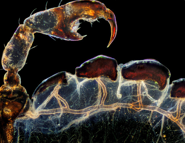
Third place: the rear leg, claw and respiratory trachea of a louse (Haematopinus suis). (Photo by Frank Reiser/Nassau Community College/Nikon Small World Photomicrography 2021)

Third place: the rear leg, claw and respiratory trachea of a louse (Haematopinus suis). (Photo by Frank Reiser/Nassau Community College/Nikon Small World Photomicrography 2021)
Details
15 Sep 2021 02:36:00
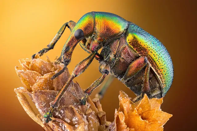
14th Place. Leaf roller weevil (Byctiscus betulae) lateral view. Image Stacking, Reflected Light 3.7X (objective lens magnification). (Photo by Özgür Kerem Bulur/Nikon Small World Photomicrography 2020)

14th Place. Leaf roller weevil (Byctiscus betulae) lateral view. Image Stacking, Reflected Light 3.7X (objective lens magnification). (Photo by Özgür Kerem Bulur/Nikon Small World Photomicrography 2020)
Details
15 Oct 2020 00:03:00
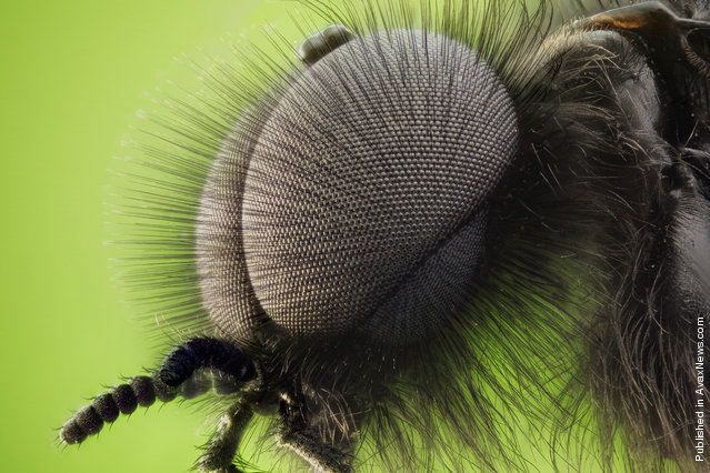
The double compound eyes of a male St. Mark's fly (Bibio marci), submitted by Dr. David Maitland from Feltwell, UK

The double compound eyes of a male St. Mark's fly (Bibio marci), submitted by Dr. David Maitland from Feltwell, UK. (Dr. David Maitland)
Details
07 Apr 2012 13:22:00
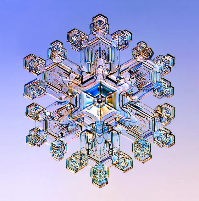
Nikon Small World Photomicrography Competition 2012. Honorable Mention. “Snow crystal, illuminated with colored lights (5x)”. (Photo by Dr. Kenneth Libbrecht, California Institute of Technology (Caltech), Department of Physics, Pasadena, California, USA)

Most people know Nikon as a purveyor of pro and consumer-grade digital cameras. But the company's expertise with optics bleeds over into related markets – it's one of the science community's major suppliers of microscopes. And each year the company asks the community to send it some of their favorite images of tiny objects. A panel of scientists and journalists have chosen the best of this past year's submissions, which Nikon has placed on its Small World site.

Photo: Honorable Mention. “Snow crystal, illuminated with colored lights (5x)”. (Photo by Dr. Kenneth Libbrecht, California Institute of Technology (Caltech), Department of Physics, Pasadena, California, USA)
Details
25 Oct 2012 13:49:00
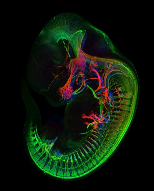
Mr. Zhong Hua, of Johns Hopkins University School of Medicine, took this image of the peripheral nerves (those outside of the brain) in an 11.5-day-old mouse embryo, magnified five times. (Photo by Zhong Hua)

Mr. Zhong Hua, of Johns Hopkins University School of Medicine, took this image of the peripheral nerves (those outside of the brain) in an 11.5-day-old mouse embryo, magnified five times. (Photo by Zhong Hua)
Details
31 Oct 2013 09:39:00