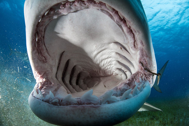
These jaw-some images show what its like to look inside the mouth of a tiger shark. Captured in amazing detail, the unique pictures show the alien-like anatomy of the shark and reveal rows upon rows of razor sharp teeth and white coloured gills. The incredible photographs were taken when a curious shark began to inspect the camera of British born photographer, Adam Hanlon, 46. After sensing electronic impulses omitted by Adams camera, the inquisitive creature began to gently mouth at the cameras housing allowing him capture the unusual perspective. (Photo by Adam Hanlon/Caters News)
29 Jan 2015 11:50:00,post received
0 comments