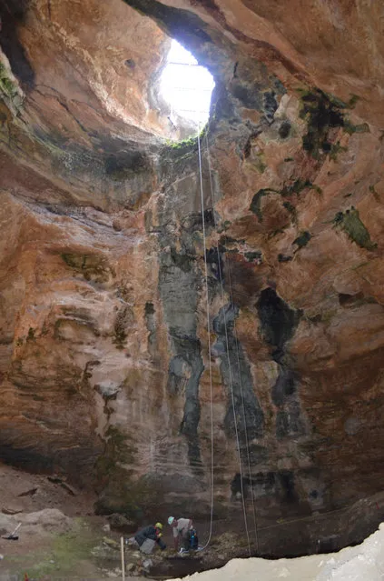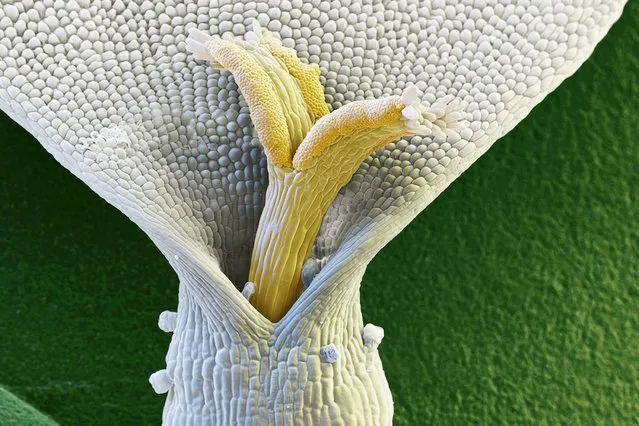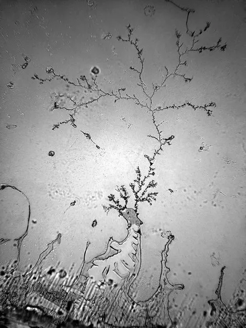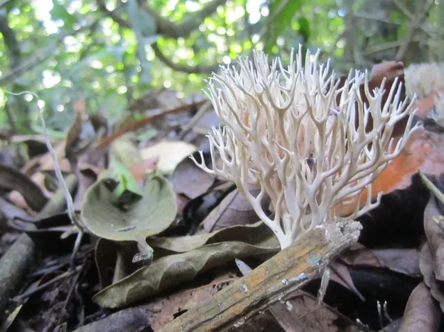
This strange coral-looking specimen is actually a mushroom. The photo, “Beautiful Destroyer”, was taken in the Panamanian tropical rainforest where the mushroom produces nitrogen, an element vital to soil health. (Photo by Sarah A. Batterman)
13 Aug 2014 09:49:00,post received
0 comments